Section of Histology and Embryology, Department of Biomedicine and Prevention, University of Rome Tor Vergata, 00133 Rome, Italy.
Department of Epidemiology, Preclinical Research and Advanced Diagnostics, National Institute for Infectious Diseases 'L. Spallanzani' IRCCS, 00149 Rome, Italy.
Cells. 2022 Apr 3;11(7):1208. doi: 10.3390/cells11071208.
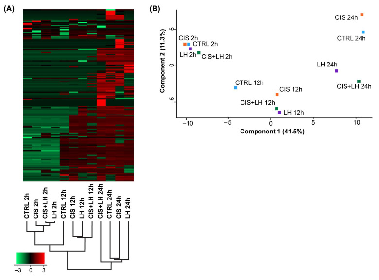
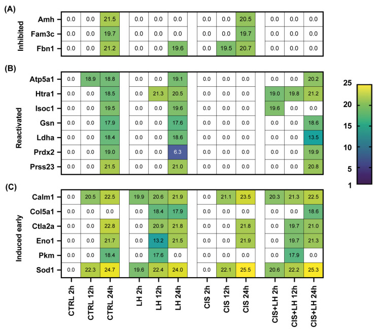
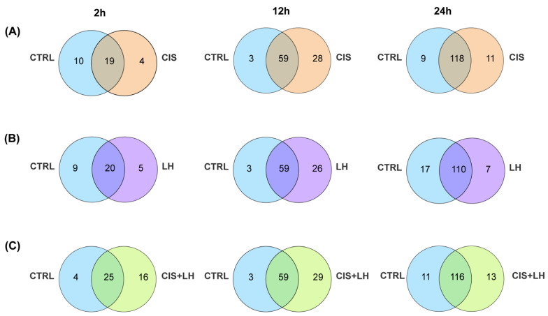
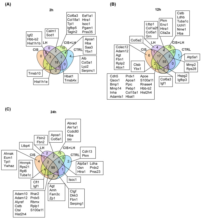
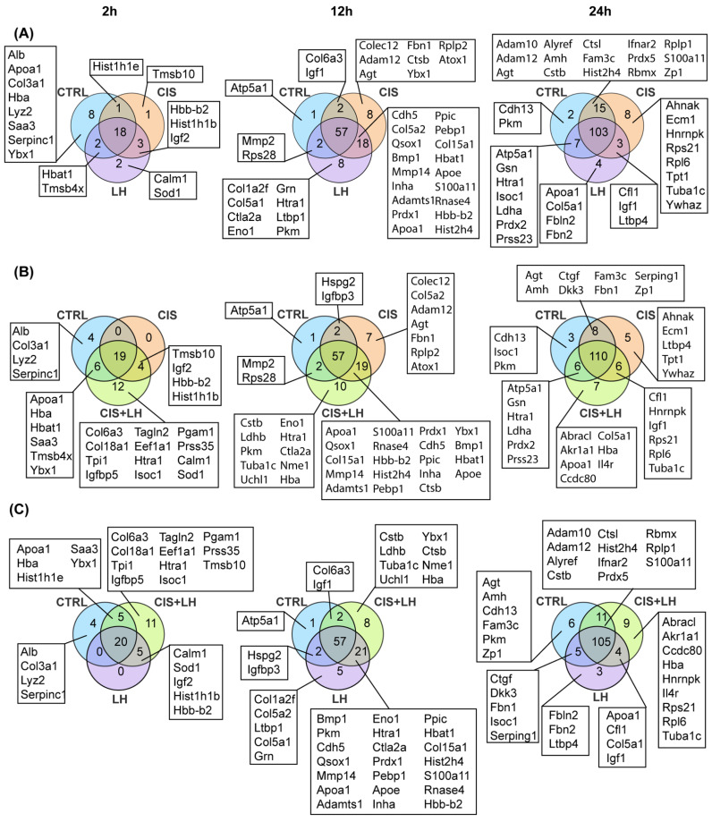
It is well known that secreted and exosomal proteins are associated with a broad range of physiological processes involving tissue homeostasis and differentiation. In the present paper, our purpose was to characterize the proteome of the culture medium in which the oocytes within the primordial/primary follicles underwent apoptosis induced by cisplatin (CIS) or were, for the most part, protected by LH against the drug. To this aim, prepubertal ovarian tissues were cultured under control and in the presence of CIS, LH, and CIS + LH. The culture media were harvested after 2, 12, and 24 h from chemotherapeutic drug treatment and analyzed by liquid chromatography-mass spectrometry (LC-MS). We found that apoptotic conditions generated by CIS in the cultured ovarian tissues and/or oocytes are reflected in distinct changes in the extracellular microenvironment in which they were cultured. These changes became evident mainly from 12 h onwards and were characterized by the inhibition or decreased release of a variety of compounds, such as the proteases Htra1 and Prss23, the antioxidants Prdx2 and Hbat1, the metabolic regulators Ldha and Pkm, and regulators of apoptotic pathways such as Tmsb4x. Altogether, these results confirm the biological relevance of the LH action on prepuberal ovaries and provide novel information about the proteins released by the ovarian tissues exposed to CIS and LH in the surrounding microenvironment. These data might represent a valuable resource for future studies aimed to clarify the effects and identify biomarkers of these compounds' action on the developing ovary.
众所周知,分泌蛋白和外泌体蛋白与广泛的生理过程有关,包括组织稳态和分化。在本文中,我们的目的是描述原始/初级卵泡中的卵母细胞在顺铂(cisplatin,cis)诱导的凋亡中或在大部分情况下由 LH 保护免受药物影响的培养介质中的蛋白质组。为此,我们在对照和存在 cis、lh 和 cis+lh 的情况下培养青春期前卵巢组织。在化疗药物处理后 2、12 和 24 小时收获培养的培养基,并通过液相色谱-质谱(LC-MS)进行分析。我们发现 cis 在培养的卵巢组织和/或卵母细胞中诱导的凋亡条件反映在它们培养的细胞外微环境中的明显变化中。这些变化主要从 12 小时开始显现,并以多种化合物的抑制或减少释放为特征,如蛋白酶 Htra1 和 Prss23、抗氧化剂 Prdx2 和 Hbat1、代谢调节剂 Ldha 和 Pkm 以及凋亡途径调节剂如 Tmsb4x。总的来说,这些结果证实了 LH 对青春期前卵巢的生物学作用,并提供了关于卵巢组织在周围微环境中暴露于 cis 和 LH 时释放的蛋白质的新信息。这些数据可能代表未来研究的有价值资源,旨在阐明这些化合物对发育中卵巢的作用的影响和鉴定生物标志物。