Iwamuro Masaya, Urata Haruo, Tanaka Takehiro, Okada Hiroyuki
Department of Gastroenterology and Hepatology, Okayama University Graduate School of Medicine, Dentistry, and Pharmaceutical Sciences, Okayama 700-8558, Japan.
Central Research Laboratory, Okayama University Medical School, Okayama 700-8558, Japan.
World J Gastrointest Pathophysiol. 2022 Mar 22;13(2):41-49. doi: 10.4291/wjgp.v13.i2.41. Epub 2022 Jan 15.
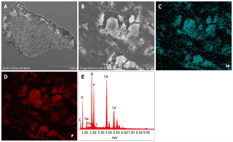
Electron microscopy has long been used in research in the fields of life sciences and materials sciences. Transmission and scanning electron microscopy and energy-dispersive X-ray spectroscopy (EDX) analyses have also been performed in the field of gastroenterology. Electron microscopy and EDX enable (1) Observation of ultrastructural differences in esophageal epithelial cells in patients with gastroesophageal reflux and eosinophilic esophagitis; (2) Detection of lanthanum deposition in the stomach and duodenum; (3) Ultrastructural and elemental analyses of enteroliths and bezoars; (4) Detection and characterization of microorganisms in the gastrointestinal tract; (5) Diagnosis of gastrointestinal tumors with neuroendocrine differentiation; and (6) Analysis of gold nanoparticles potentially used in endoscopic photodynamic therapy. This review aims to foster a better understanding of electron microscopy applications by reviewing relevant clinical studies, basic research findings, and the state of current research carried out in gastroenterology science.
电子显微镜长期以来一直应用于生命科学和材料科学领域的研究。透射电子显微镜、扫描电子显微镜以及能量色散X射线光谱(EDX)分析也已在胃肠病学领域开展。电子显微镜和EDX能够:(1)观察胃食管反流和嗜酸性食管炎患者食管上皮细胞的超微结构差异;(2)检测胃和十二指肠中的镧沉积;(3)对肠石和胃石进行超微结构和元素分析;(4)检测和鉴定胃肠道中的微生物;(5)诊断具有神经内分泌分化的胃肠道肿瘤;以及(6)分析可能用于内镜光动力治疗的金纳米颗粒。本综述旨在通过回顾相关临床研究、基础研究结果以及胃肠病学领域当前的研究状况,促进对电子显微镜应用的更好理解。