Department of Precision and Microsystems Engineering, Delft University of Technology, Mekelweg 2, 2628 CD Delft, The Netherlands.
Holland Proton Therapy Center (HollandPTC), Huismansingel 4, 2629 JH Delft, The Netherlands.
ACS Appl Mater Interfaces. 2022 May 11;14(18):20778-20789. doi: 10.1021/acsami.2c03706. Epub 2022 Apr 20.
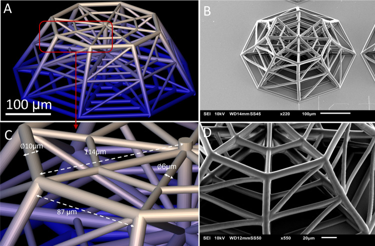
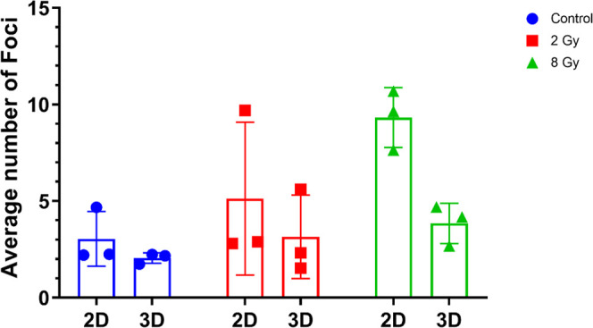
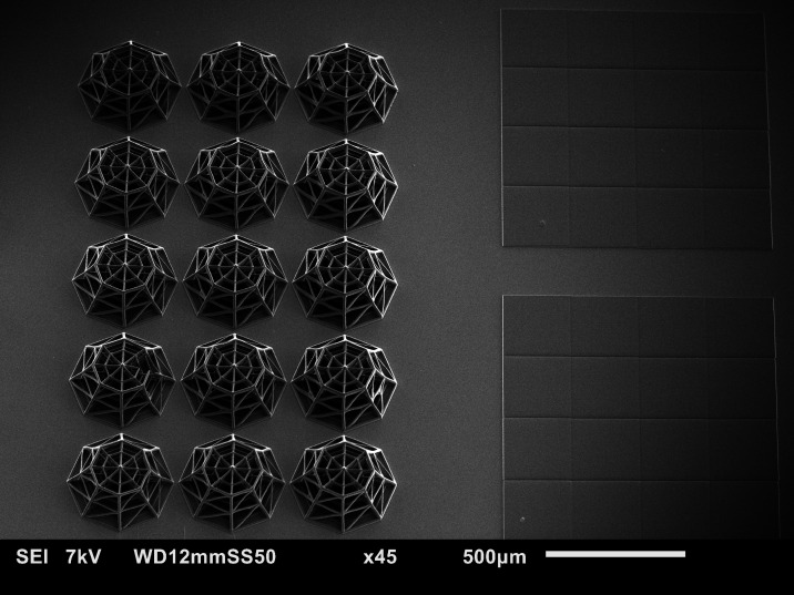
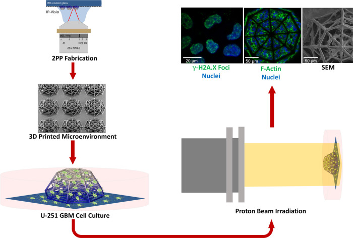
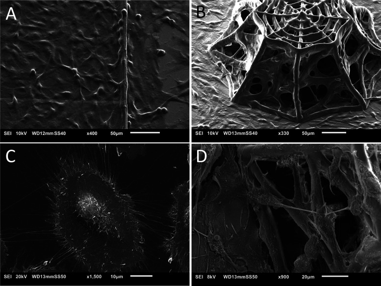
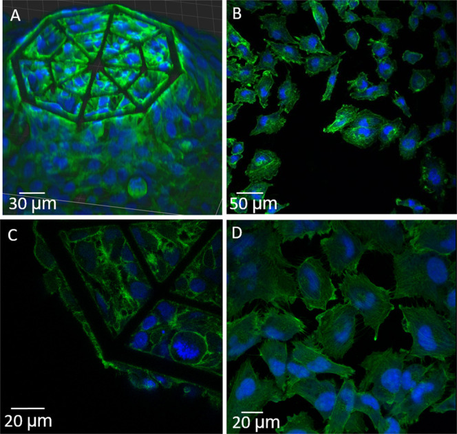
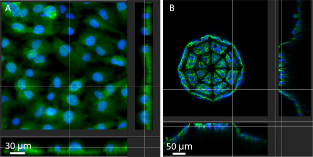
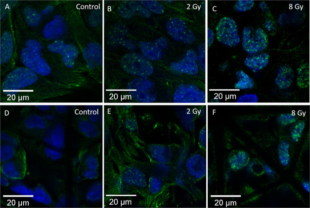
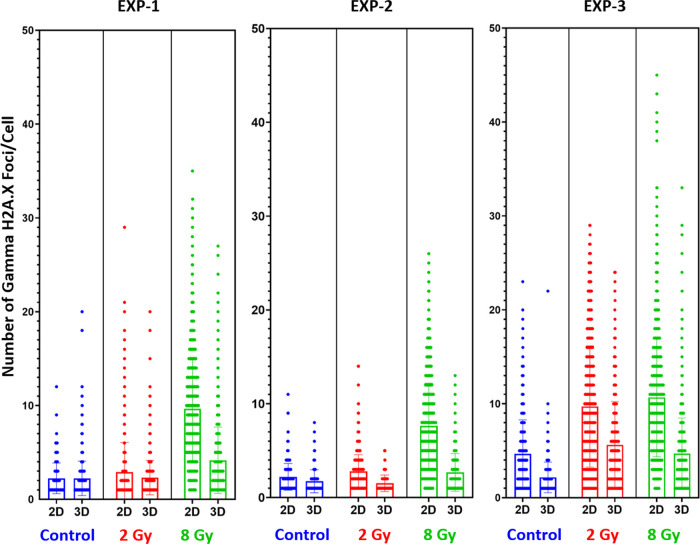
Glioblastoma (GBM) is a devastating cancer of the brain with an extremely poor prognosis. For this reason, besides clinical and preclinical studies, novel models for the assessment of cancer response to drugs and radiation are being developed. In such context, three-dimensional (3D)-engineered cellular microenvironments, compared to unrealistic two-dimensional (2D) monolayer cell culture, provide a model closer to the configuration. Concerning cancer treatment, while X-ray radiotherapy and chemotherapy remain the current standard, proton beam therapy is an appealing alternative as protons can be efficiently targeted to destroy cancer cells while sparing the surrounding healthy tissue. However, despite the treatment's compelling biological and medical rationale, little is known about the effects of protons on GBM at the cellular level. In this work, we designed novel 3D-engineered scaffolds inspired by the geometry of brain blood vessels, which cover a vital role in the colonization mechanisms of GBM cells. The architectures were fabricated by two-photon polymerization (2PP), cultured with U-251 GBM cells and integrated for the first time in the context of proton radiation experiments to assess their response to treatment. We employed Gamma H2A.X as a fluorescent biomarker to identify the DNA damage induced in the cells by proton beams. The results show a higher DNA double-strand breakage in 2D cell monolayers as compared to cells cultured in 3D. The discrepancy in terms of proton radiation response could indicate a difference in the radioresistance of the GBM cells or in the rate of repair kinetics between 2D cell monolayers and 3D cell networks. Thus, these biomimetic-engineered 3D scaffolds pave the way for the realization of a benchmark tool that can be used to routinely assess the effects of proton therapy on 3D GBM cell networks and other types of cancer cells.
胶质母细胞瘤(GBM)是一种具有极差预后的毁灭性脑部癌症。因此,除了临床和临床前研究外,还在开发新的模型来评估癌症对药物和辐射的反应。在这种情况下,与不切实际的二维(2D)单层细胞培养相比,三维(3D)工程细胞微环境为更接近实际情况的模型提供了可能性。关于癌症治疗,虽然 X 射线放疗和化疗仍然是目前的标准,但质子束治疗作为一种有吸引力的替代方法,因为质子可以有效地靶向杀死癌细胞,同时保护周围的健康组织。然而,尽管这种治疗具有引人注目的生物学和医学原理,但人们对质子束在细胞水平上对 GBM 的影响知之甚少。在这项工作中,我们设计了受脑血管几何形状启发的新型 3D 工程支架,这些支架在 GBM 细胞定植机制中起着重要作用。这些结构是通过双光子聚合(2PP)制造的,与 U-251 GBM 细胞共培养,并首次整合到质子辐射实验中,以评估它们对治疗的反应。我们采用γH2A.X 作为荧光生物标志物来识别质子束在细胞中诱导的 DNA 损伤。结果表明,与在 3D 中培养的细胞相比,2D 细胞单层中的 DNA 双链断裂更多。质子辐射反应方面的差异可能表明 GBM 细胞的放射抗性或 2D 细胞单层和 3D 细胞网络之间的修复动力学速率存在差异。因此,这些仿生工程 3D 支架为实现基准工具铺平了道路,该工具可用于常规评估质子治疗对 3D GBM 细胞网络和其他类型癌细胞的影响。