Institute of Metabolism and Systems Research, Birmingham Medical School, University of Birmingham, Birmingham, UK.
Biosciences Institute, The Medical School, Newcastle University, Newcastle upon Tyne, UK.
Nutr Diabetes. 2022 Apr 20;12(1):22. doi: 10.1038/s41387-022-00199-y.
Rodent and human β-cells are differentially susceptible to the "lipotoxic" effects of long-chain saturated fatty acids (LC-SFA) but the factors accounting for this are unclear. Here, we have studied the intracellular disposition of the LC-SFA palmitate in human vs rodent β-cells and present data that reveal new insights into the factors regulating β-cell lipotoxicity.
The subcellular distribution of the LC-SFA palmitate was studied in rodent (INS-1E and INS-1 823/13 cells) and human (EndoC-βH1) β-cells using confocal fluorescence and electron microscopy (EM). Protein expression was assessed by Western blotting and cell viability, by vital dye staining.
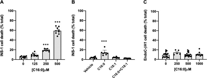
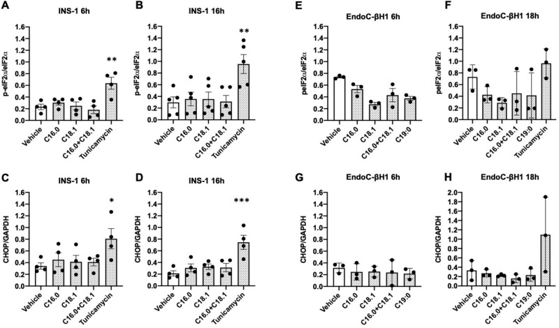
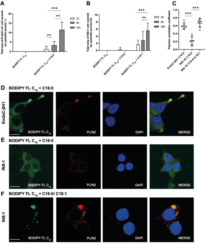
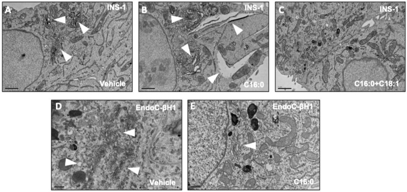
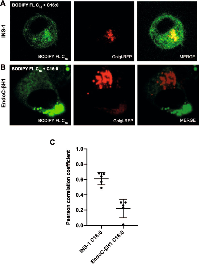
Exposure of INS-1 cells to palmitate for 24 h led to loss of viability, whereas EndoC-βH1 cells remained viable even after 72 h of treatment with a high concentration (1 mM) of palmitate. Use of the fluorescent palmitate analogue BODIPY FL C revealed an early localisation of the LC-SFA to the Golgi apparatus in INS-1 cells and this correlated with distention of intracellular membranes, visualised under the EM. Despite this, the PERK-dependent ER stress pathway was not activated under these conditions. By contrast, BODIPY FL C did not accumulate in the Golgi apparatus in EndoC-βH1 cells but, rather, co-localised with the lipid droplet-associated protein, PLIN2, suggesting preferential routing into lipid droplets. When INS-1 cells were treated with a combination of palmitate plus oleate, the toxic effects of palmitate were attenuated and BODIPY FL C localised primarily with PLIN2 but not with a Golgi marker.
In rodent β-cells, palmitate accumulates in the Golgi apparatus at early time points whereas, in EndoC- βH1 cells, it is routed preferentially into lipid droplets. This may account for the differential sensitivity of rodent vs human β-cells to "lipotoxicity" since manoeuvres leading to the incorporation of palmitate into lipid droplets is associated with the maintenance of cell viability in both cell types.
长链饱和脂肪酸(LC-SFA)的“脂毒性”对啮齿动物和人类β细胞的影响不同,但造成这种差异的因素尚不清楚。在这里,我们研究了 LC-SFA 棕榈酸在人和啮齿动物β细胞中的细胞内分布,并提供了新的见解,揭示了调节β细胞脂毒性的因素。
使用共聚焦荧光和电子显微镜(EM)研究 LC-SFA 棕榈酸在啮齿动物(INS-1E 和 INS-1 823/13 细胞)和人(EndoC-βH1)β细胞中的亚细胞分布。通过 Western blot 评估蛋白表达,通过活染料染色评估细胞活力。
暴露于棕榈酸 24 小时可导致 INS-1 细胞活力丧失,而 EndoC-βH1 细胞即使在高浓度(1mM)棕榈酸处理 72 小时后仍保持活力。使用荧光棕榈酸类似物 BODIPY FL C 显示 LC-SFA 早期定位于 INS-1 细胞的高尔基体,这与 EM 下观察到的细胞内膜扩张相关。尽管如此,在这些条件下,PERK 依赖性内质网应激途径并未被激活。相比之下,BODIPY FL C 不会在 EndoC-βH1 细胞的高尔基体中积累,而是与脂滴相关蛋白 PLIN2 共定位,表明优先定向进入脂滴。当 INS-1 细胞用棕榈酸加油酸处理时,棕榈酸的毒性作用减弱,BODIPY FL C 主要与 PLIN2 共定位,而不是与高尔基体标记物共定位。
在啮齿动物β细胞中,棕榈酸在早期时间点积聚在高尔基体中,而在 EndoC-βH1 细胞中,它优先定向进入脂滴。这可能解释了啮齿动物和人类β细胞对“脂毒性”的敏感性差异,因为导致棕榈酸掺入脂滴的操作与两种细胞类型的细胞活力维持有关。