Department of Ophthalmology, Affiliated Hospital of Inner Mongolia Minzu University, Tongliao, Inner Mongolia, P.R. China.
Department of Ophthalmology, Shanxi Eye Hospital, Taiyuan, Shanxi, P.R. China.
Transl Vis Sci Technol. 2022 Apr 1;11(4):20. doi: 10.1167/tvst.11.4.20.
To evaluate microvascular abnormalities in the macula and peripapillary area in diabetic patients without clinical signs of diabetic retinopathy (DR) and compare them with healthy control eyes, using optical coherence tomography angiography (OCTA).
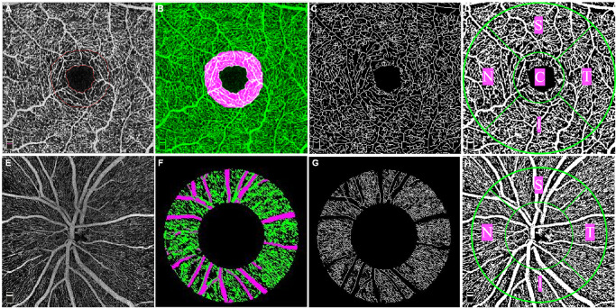
A prospective study was performed of 49 eyes from 49 diabetic patients without clinical signs of DR and a control group of 52 eyes from 52 healthy normal individuals. The 3 × 3 mm macular scans and 4.5 × 4.5 mm optic disc scans were obtained with the OCTA RTVue-XR Avanti system. Angiograms from the superficial capillary plexus, the deep capillary plexus of the macula scans, and radial peripapillary capillary plexus of the optic disc scans were analyzed with MATLAB. Multivariate binary logistic regression and the least absolute shrinkage and selection operator (LASSO) regression were used to select ideal parameters that distinguish diabetic eyes without DR from normal eyes. A receiver operating characteristic (ROC) curve was generated, and sensitivity and specificity were calculated.
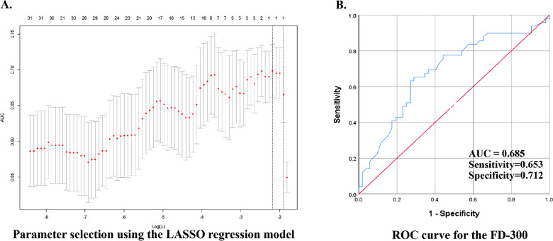
Our final model identified FD-300 (foveal vessel density in a 300-µm-wide region around foveal avascular zone) as the only parameter selected by both the LASSO regression and the final multivariate logistic regression model that significantly differentiates diabetic eyes without clinical signs of DR from healthy normal eyes. The area under the ROC curve of FD-300 was 0.685, and sensitivity and specificity were 65.3% and 71.2%, respectively.
Quantitative evaluation of retinal microvascular abnormalities using OCTA identified FD-300 as a useful biomarker versus the other macular and peripapillary OCTA metrics in the early detection of preclinical diabetic retinal abnormalities.
OCTA may be useful in detecting early retinal microvascular abnormalities in diabetic patients before the clinical findings of DR become visible.
利用光相干断层扫描血管造影术(OCTA)评估无临床糖尿病视网膜病变(DR)征象的糖尿病患者黄斑区和视盘周围区域的微血管异常,并与健康对照组进行比较。
对 49 例无临床 DR 征象的糖尿病患者的 49 只眼和 52 例健康正常人的 52 只眼进行前瞻性研究。使用 OCTA RTVue-XR Avanti 系统获取 3×3mm 黄斑扫描和 4.5×4.5mm 视盘扫描。使用 MATLAB 分析来自浅层毛细血管丛、黄斑扫描深层毛细血管丛和视盘扫描放射状周边毛细血管丛的血管造影图。采用多元二项逻辑回归和最小绝对收缩和选择算子(LASSO)回归选择理想的参数来区分无 DR 的糖尿病眼和正常眼。生成受试者工作特征(ROC)曲线,并计算灵敏度和特异性。
我们的最终模型确定 FD-300(黄斑无血管区周围 300μm 宽区域的中心凹血管密度)是 LASSO 回归和最终多元逻辑回归模型均选择的唯一参数,可显著区分无临床 DR 征象的糖尿病眼和正常眼。FD-300 的 ROC 曲线下面积为 0.685,灵敏度和特异性分别为 65.3%和 71.2%。
OCTA 定量评估视网膜微血管异常可识别 FD-300,作为一种有用的生物标志物,与其他黄斑和视盘 OCTA 指标相比,可在临床发现 DR 之前更早地检测到糖尿病视网膜早期异常。