• 文献检索
  • 文档翻译
  • 深度研究
  • 学术资讯
  • Suppr Zotero 插件Zotero 插件
  • 邀请有礼
  • 套餐&价格
  • 历史记录
应用&插件
Suppr Zotero 插件Zotero 插件浏览器插件Mac 客户端Windows 客户端微信小程序
定价
高级版会员购买积分包购买API积分包
服务
文献检索文档翻译深度研究API 文档MCP 服务
关于我们
关于 Suppr公司介绍联系我们用户协议隐私条款
关注我们

Suppr 超能文献

核心技术专利:CN118964589B侵权必究
粤ICP备2023148730 号-1Suppr @ 2026

文献检索

告别复杂PubMed语法,用中文像聊天一样搜索,搜遍4000万医学文献。AI智能推荐,让科研检索更轻松。

立即免费搜索

文件翻译

保留排版,准确专业,支持PDF/Word/PPT等文件格式,支持 12+语言互译。

免费翻译文档

深度研究

AI帮你快速写综述,25分钟生成高质量综述,智能提取关键信息,辅助科研写作。

立即免费体验

应激心脏中NOTCH1信号通路的抑制可限制纤维化并促进非心肌细胞向心肌细胞命运的募集。

Inhibition of the NOTCH1 Pathway in the Stressed Heart Limits Fibrosis and Promotes Recruitment of Non-Myocyte Cells into the Cardiomyocyte Fate.

作者信息

Nemir Mohamed, Kay Maryam, Maison Damien, Berthonneche Corinne, Sarre Alexandre, Plaisance Isabelle, Pedrazzini Thierry

机构信息

Experimental Cardiology Unit, Division of Cardiology, Department of Cardiovascular Medicine, University of Lausanne Medical School, 1011 Lausanne, Switzerland.

Cardiovascular Assessment Facility, University of Lausanne, 1011 Lausanne, Switzerland.

出版信息

J Cardiovasc Dev Dis. 2022 Apr 7;9(4):111. doi: 10.3390/jcdd9040111.

DOI:10.3390/jcdd9040111
PMID:35448087
原文链接:https://pmc.ncbi.nlm.nih.gov/articles/PMC9024539/
Abstract

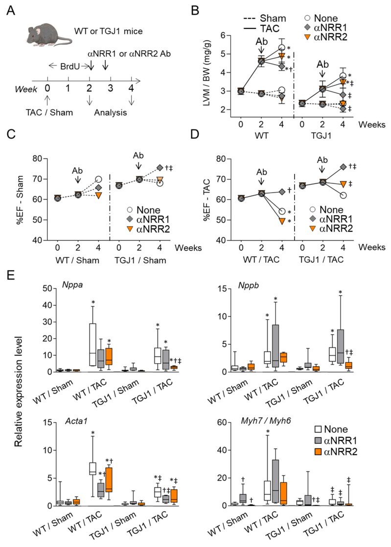
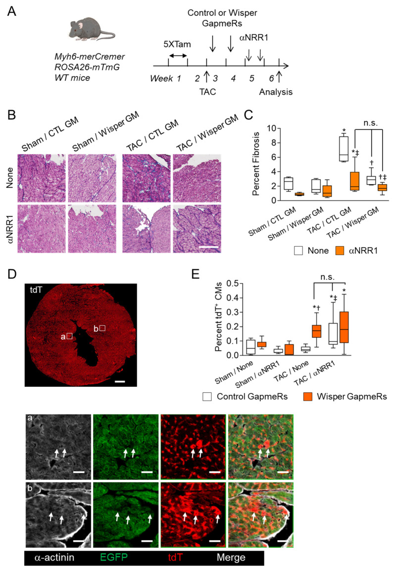
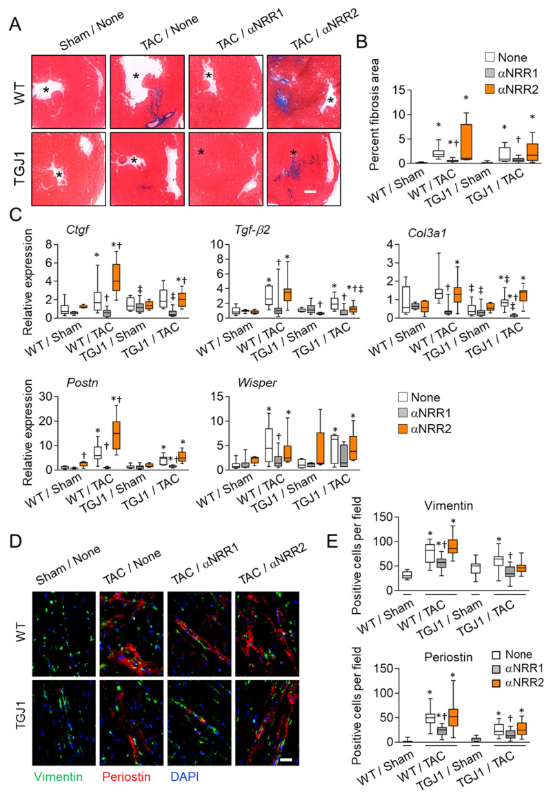
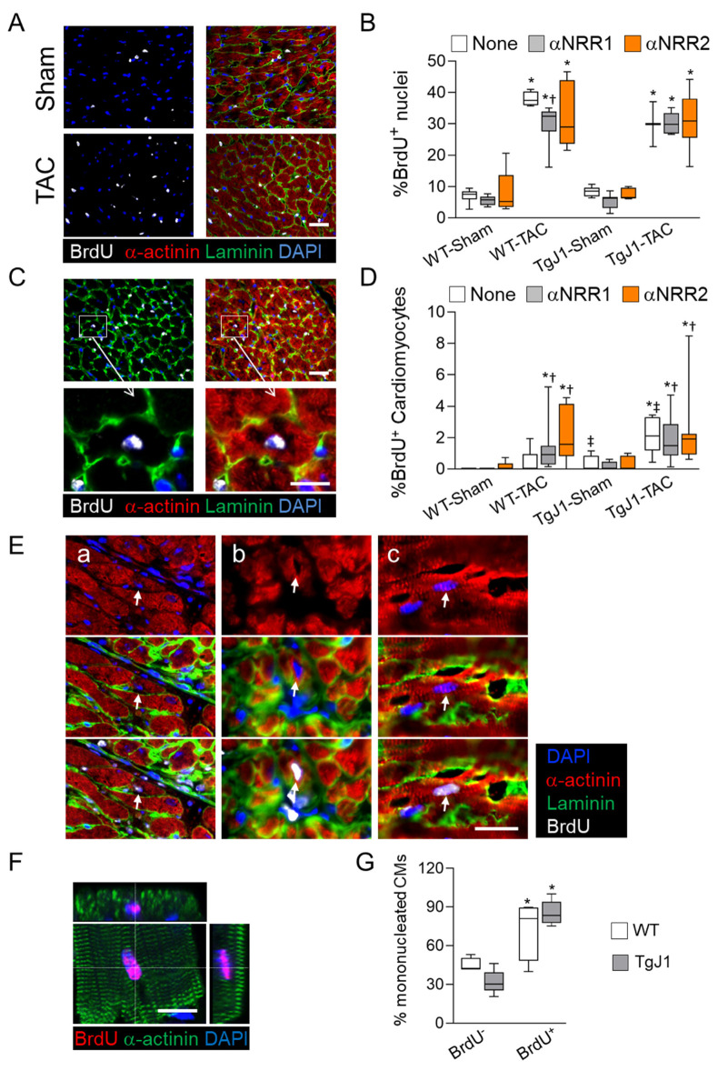
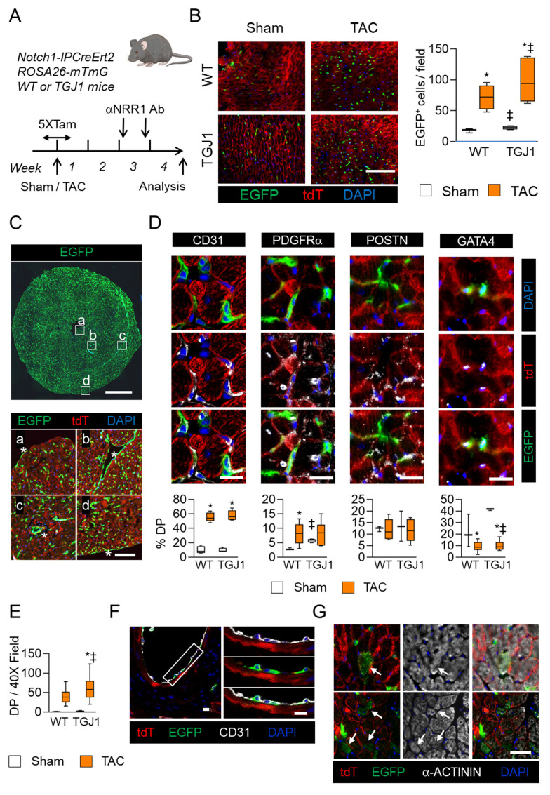
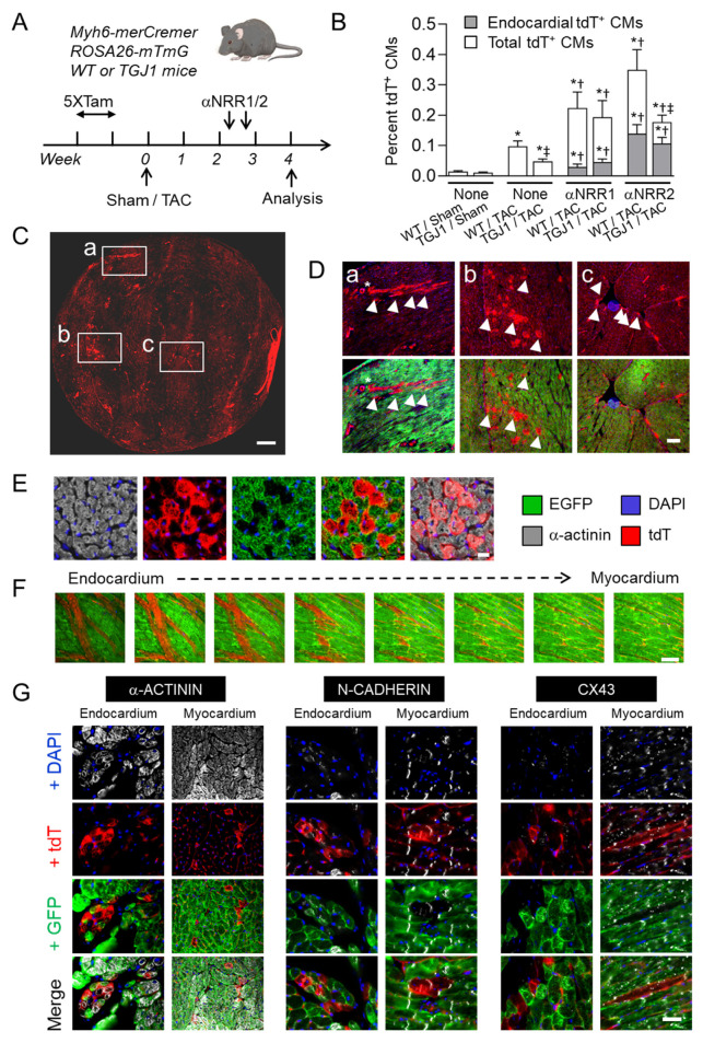
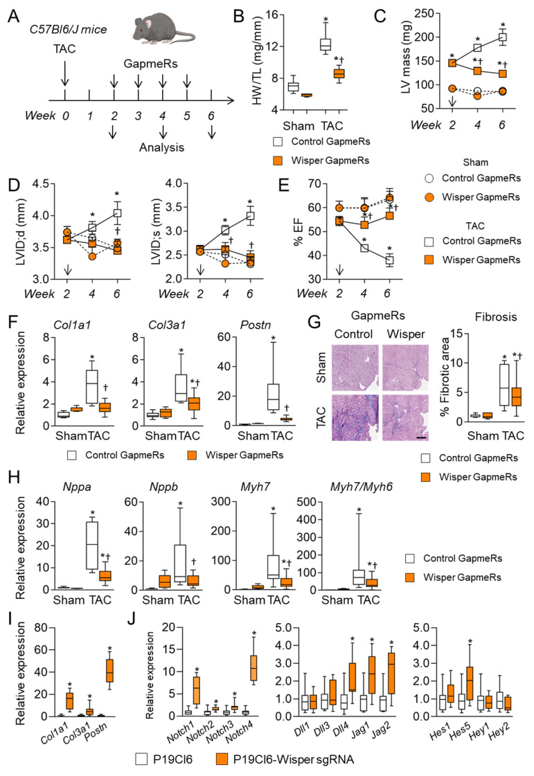
Cardiac pathologies lead to an acute or gradual loss of cardiomyocytes. Because of the limited regenerative capacity of the mammalian heart, cardiomyocytes are only replaced by fibrotic tissue. Excessive fibrosis contributes to the deterioration of cardiac function and the transition to heart failure, which is the leading cause of morbidity and mortality worldwide. Currently, no treatments can promote replenishment of the injured heart with newly formed cardiomyocytes. In this context, regenerative strategies explore the possibility to promote recovery through induction of cardiomyocyte production from pre-existing cardiomyocytes. On the other hand, cardiac non-myocyte cells can be directly reprogrammed into induced cardiac precursor cells and cardiomyocytes, suggesting that these cells could be exploited to produce cardiomyocytes in vivo. Here, we provide evidence that the sequential activation and inhibition of the NOTCH1 signaling pathway in the stressed heart decreases fibrosis and improves cardiac function in the stressed heart. This is accompanied by the emergence of new cardiomyocytes from non-myocyte origin. Overall, our data show how a developmental pathway such as the NOTCH pathway can be manipulated to provide therapeutic benefit in the damaged heart.

摘要

心脏疾病会导致心肌细胞急性或逐渐丧失。由于哺乳动物心脏的再生能力有限,心肌细胞只能被纤维组织替代。过度纤维化会导致心脏功能恶化并发展为心力衰竭,而心力衰竭是全球发病和死亡的主要原因。目前,尚无治疗方法能够促进损伤的心脏重新生成新的心肌细胞。在此背景下,再生策略探索了通过诱导已有心肌细胞产生新的心肌细胞来促进恢复的可能性。另一方面,心脏非心肌细胞可直接重编程为诱导性心脏前体细胞和心肌细胞,这表明这些细胞可用于在体内产生心肌细胞。在此,我们提供证据表明,在应激心脏中依次激活和抑制NOTCH1信号通路可减少纤维化并改善应激心脏的功能。同时,会出现源自非心肌细胞的新的心肌细胞。总体而言,我们的数据表明,像NOTCH通路这样的发育途径可以被操控以对受损心脏提供治疗益处。

https://cdn.ncbi.nlm.nih.gov/pmc/blobs/1e5b/9024539/4521c9600dc9/jcdd-09-00111-g007.jpg
https://cdn.ncbi.nlm.nih.gov/pmc/blobs/1e5b/9024539/b726820a5d10/jcdd-09-00111-g001.jpg
https://cdn.ncbi.nlm.nih.gov/pmc/blobs/1e5b/9024539/b3192755b84a/jcdd-09-00111-g002.jpg
https://cdn.ncbi.nlm.nih.gov/pmc/blobs/1e5b/9024539/a7692bae93f8/jcdd-09-00111-g003.jpg
https://cdn.ncbi.nlm.nih.gov/pmc/blobs/1e5b/9024539/3fadc9af03a1/jcdd-09-00111-g004.jpg
https://cdn.ncbi.nlm.nih.gov/pmc/blobs/1e5b/9024539/fa3e2c33a2ca/jcdd-09-00111-g005.jpg
https://cdn.ncbi.nlm.nih.gov/pmc/blobs/1e5b/9024539/4770b318d58b/jcdd-09-00111-g006.jpg
https://cdn.ncbi.nlm.nih.gov/pmc/blobs/1e5b/9024539/4521c9600dc9/jcdd-09-00111-g007.jpg

相似文献

1
Inhibition of the NOTCH1 Pathway in the Stressed Heart Limits Fibrosis and Promotes Recruitment of Non-Myocyte Cells into the Cardiomyocyte Fate.应激心脏中NOTCH1信号通路的抑制可限制纤维化并促进非心肌细胞向心肌细胞命运的募集。
J Cardiovasc Dev Dis. 2022 Apr 7;9(4):111. doi: 10.3390/jcdd9040111.
2
The Notch pathway controls fibrotic and regenerative repair in the adult heart.Notch信号通路控制成年心脏的纤维化和再生修复。
Eur Heart J. 2014 Aug 21;35(32):2174-85. doi: 10.1093/eurheartj/ehs269. Epub 2012 Nov 19.
3
Control of the adaptive response of the heart to stress via the Notch1 receptor pathway.通过Notch1受体途径控制心脏对压力的适应性反应。
J Exp Med. 2008 Dec 22;205(13):3173-85. doi: 10.1084/jem.20081427. Epub 2008 Dec 8.
4
Jagged1 intracellular domain-mediated inhibition of Notch1 signalling regulates cardiac homeostasis in the postnatal heart.锯齿蛋白1细胞内结构域介导的Notch1信号抑制调节出生后心脏的心脏稳态。
Cardiovasc Res. 2015 Oct 1;108(1):74-86. doi: 10.1093/cvr/cvv209. Epub 2015 Aug 6.
5
Cardiac Fibroblasts and Myocardial Regeneration.心脏成纤维细胞与心肌再生
Front Bioeng Biotechnol. 2021 Mar 25;9:599928. doi: 10.3389/fbioe.2021.599928. eCollection 2021.
6
Redox Regulation of Heart Regeneration: An Evolutionary Tradeoff.心脏再生的氧化还原调节:一种进化权衡。
Front Cell Dev Biol. 2016 Dec 15;4:137. doi: 10.3389/fcell.2016.00137. eCollection 2016.
7
Is zebrafish heart regeneration "complete"? Lineage-restricted cardiomyocytes proliferate to pre-injury numbers but some fail to differentiate in fibrotic hearts.斑马鱼的心脏再生是“完全的”吗?谱系限制的心肌细胞增殖到损伤前的数量,但在纤维化的心脏中有些未能分化。
Dev Biol. 2021 Mar;471:106-118. doi: 10.1016/j.ydbio.2020.12.004. Epub 2020 Dec 10.
8
Leading progress in heart regeneration and repair.引领心脏再生和修复领域的进展。
Curr Opin Cell Biol. 2019 Dec;61:79-85. doi: 10.1016/j.ceb.2019.07.005. Epub 2019 Aug 10.
9
Cardiomyocyte Proliferation as a Source of New Myocyte Development in the Adult Heart.成体心脏中心肌细胞增殖作为新心肌细胞发育的来源。
Int J Mol Sci. 2021 Jul 21;22(15):7764. doi: 10.3390/ijms22157764.
10
Cardiac myocyte p38α kinase regulates angiogenesis via myocyte-endothelial cell cross-talk during stress-induced remodeling in the heart.在心脏应激诱导重塑过程中,心肌细胞p38α激酶通过心肌细胞与内皮细胞的相互作用调节血管生成。
J Biol Chem. 2017 Aug 4;292(31):12787-12800. doi: 10.1074/jbc.M117.784553. Epub 2017 Jun 21.

引用本文的文献

1
Research Advances in Myocardial Infarction Repair and Cardiac Regenerative Medicine via the Notch Signaling Pathway.通过Notch信号通路进行心肌梗死修复及心脏再生医学的研究进展
Rev Cardiovasc Med. 2025 Mar 19;26(3):26587. doi: 10.31083/RCM26587. eCollection 2025 Mar.
2
Inhibiting AGTR1 reduces AML burden and protects the heart from cardiotoxicity in mouse models.抑制 AGTR1 可减轻 AML 负担并保护心脏免受心脏毒性的影响。
Sci Transl Med. 2024 Jun 19;16(752):eadl5931. doi: 10.1126/scitranslmed.adl5931.
3
Effects of maternal hypothyroidism on postnatal cardiomyocyte proliferation and cardiac disease responses of the progeny.

本文引用的文献

1
Disruption of myofibroblastic Notch signaling attenuates liver fibrosis by modulating fibrosis progression and regression.肌成纤维细胞 Notch 信号通路的破坏通过调节肝纤维化的进展和消退来减轻肝纤维化。
Int J Biol Sci. 2021 May 27;17(9):2135-2146. doi: 10.7150/ijbs.60056. eCollection 2021.
2
Cardiac fibrosis: potential therapeutic targets.心脏纤维化:潜在的治疗靶点。
Transl Res. 2019 Jul;209:121-137. doi: 10.1016/j.trsl.2019.03.001. Epub 2019 Mar 9.
3
Reassessing endothelial-to-mesenchymal transition in cardiovascular diseases.
母体甲状腺功能减退症对子代心肌细胞增殖和心脏疾病反应的影响。
Am J Physiol Heart Circ Physiol. 2023 Oct 1;325(4):H702-H719. doi: 10.1152/ajpheart.00320.2023. Epub 2023 Aug 4.
4
Direct Reprogramming of Resident Non-Myocyte Cells and Its Potential for In Vivo Cardiac Regeneration.常驻非心肌细胞的直接重编程及其在体内心脏再生中的潜力。
Cells. 2023 Apr 15;12(8):1166. doi: 10.3390/cells12081166.
重新评估心血管疾病中的内皮细胞向间充质细胞转化。
Nat Rev Cardiol. 2018 Aug;15(8):445-456. doi: 10.1038/s41569-018-0023-y.
4
Cardiomyocyte Lineage Specification in Adult Human Cardiac Precursor Cells Via Modulation of Enhancer-Associated Long Noncoding RNA Expression.通过调节增强子相关长链非编码RNA表达实现成人心脏前体细胞中的心肌细胞谱系特化
JACC Basic Transl Sci. 2016 Oct;1(6):472-493. doi: 10.1016/j.jacbts.2016.06.008.
5
Deletion of delta-like 1 homologue accelerates fibroblast-myofibroblast differentiation and induces myocardial fibrosis.Delta-like 1 同源物缺失加速成纤维细胞-肌成纤维细胞分化并诱导心肌纤维化。
Eur Heart J. 2019 Mar 21;40(12):967-978. doi: 10.1093/eurheartj/ehy188.
6
Fibroblast-specific TGF-β-Smad2/3 signaling underlies cardiac fibrosis.成纤维细胞特异性转化生长因子-β- Smad2/3信号传导是心脏纤维化的基础。
J Clin Invest. 2017 Oct 2;127(10):3770-3783. doi: 10.1172/JCI94753. Epub 2017 Sep 11.
7
Potential role of the Jagged1/Notch1 signaling pathway in the endothelial-myofibroblast transition during BLM-induced pulmonary fibrosis.Jagged1/Notch1信号通路在博来霉素诱导的肺纤维化过程中内皮细胞-肌成纤维细胞转分化中的潜在作用
J Cell Physiol. 2018 Mar;233(3):2451-2463. doi: 10.1002/jcp.26122. Epub 2017 Sep 7.
8
Cardiomyocyte Regeneration: A Consensus Statement.心肌细胞再生:一份共识声明。
Circulation. 2017 Aug 15;136(7):680-686. doi: 10.1161/CIRCULATIONAHA.117.029343. Epub 2017 Jul 6.
9
Artesunate ameliorates lung fibrosis via inhibiting the Notch signaling pathway.青蒿琥酯通过抑制Notch信号通路改善肺纤维化。
Exp Ther Med. 2017 Jul;14(1):561-566. doi: 10.3892/etm.2017.4573. Epub 2017 Jun 8.
10
The long noncoding RNA controls cardiac fibrosis and remodeling.长链非编码RNA控制心脏纤维化和重塑。
Sci Transl Med. 2017 Jun 21;9(395). doi: 10.1126/scitranslmed.aai9118.