Holzgreve Adrien, Pötter Dennis, Brendel Matthias, Orth Michael, Weidner Lorraine, Gold Lukas, Kirchner Maximilian A, Bartos Laura M, Unterrainer Lena M, Unterrainer Marcus, Steiger Katja, von Baumgarten Louisa, Niyazi Maximilian, Belka Claus, Bartenstein Peter, Riemenschneider Markus J, Lauber Kirsten, Albert Nathalie L
Department of Nuclear Medicine, University Hospital, Ludwig Maximilian University of Munich (LMU Munich), 81377 Munich, Germany.
Department of Radiation Oncology, University Hospital, Ludwig Maximilian University of Munich (LMU Munich), 81377 Munich, Germany.
Biomedicines. 2022 Mar 22;10(4):738. doi: 10.3390/biomedicines10040738.
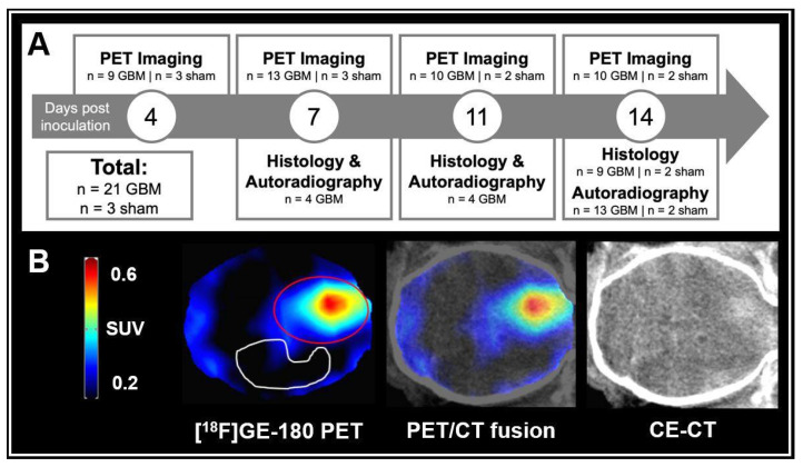
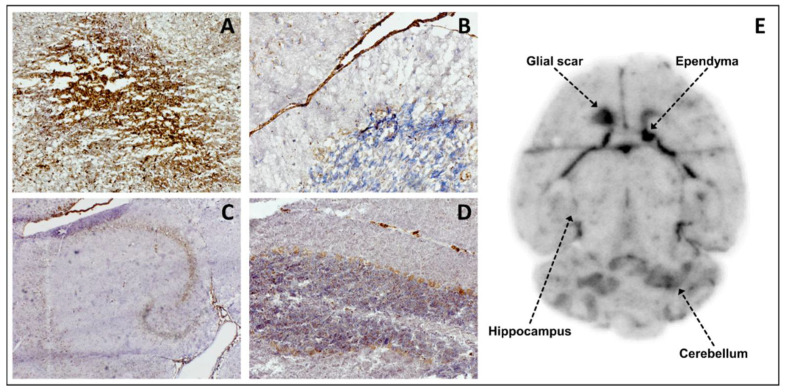
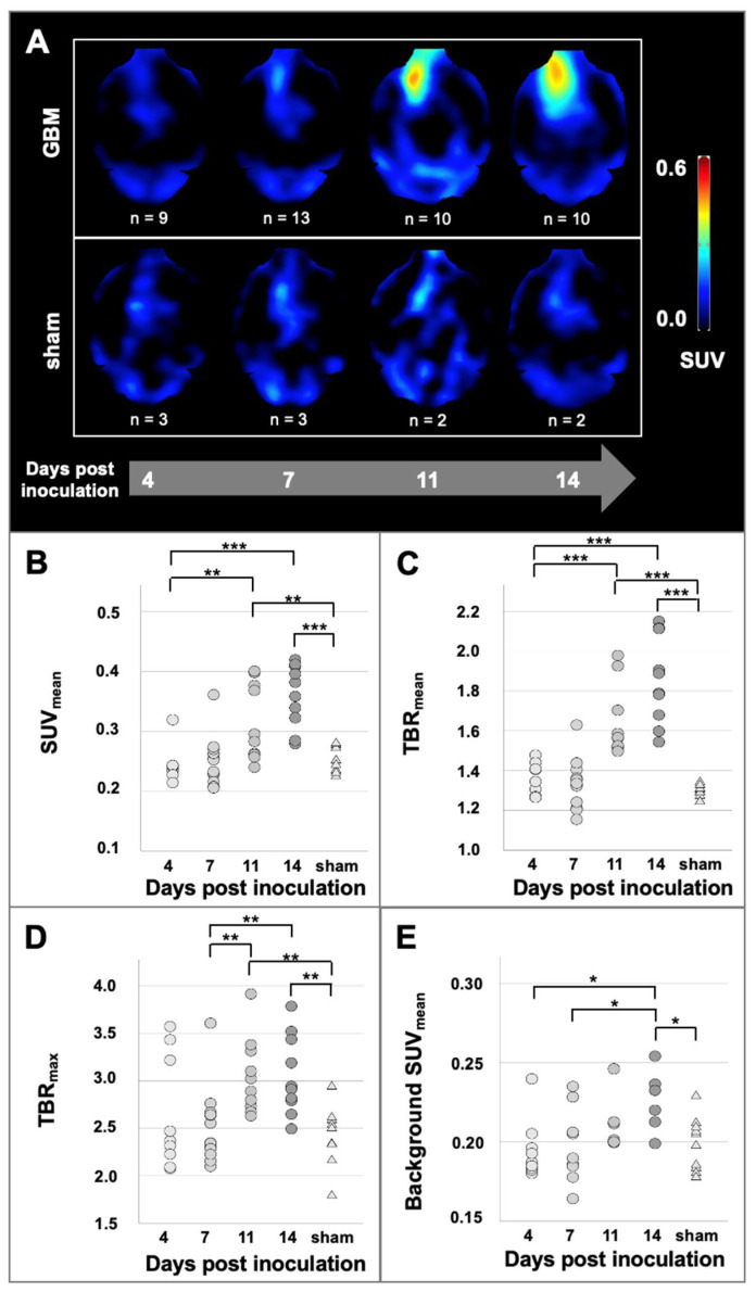
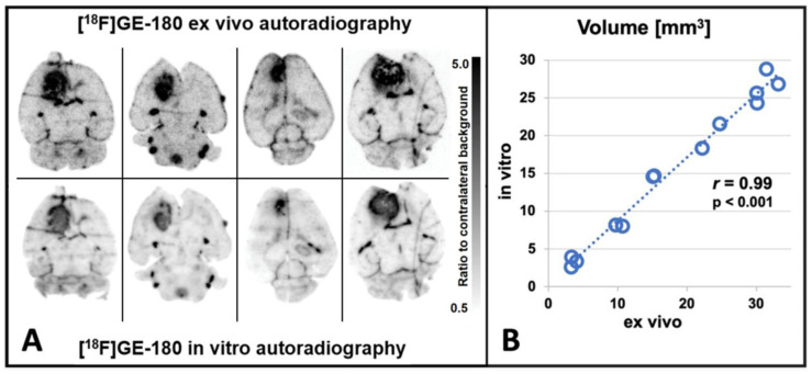
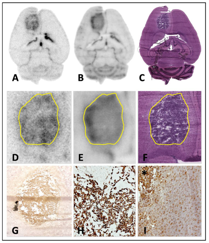
The 18 kDa translocator protein (TSPO) is increasingly recognized as an interesting target for the imaging of glioblastoma (GBM). Here, we investigated TSPO PET imaging and autoradiography in the frequently used GL261 glioblastoma mouse model and aimed to generate insights into the temporal evolution of TSPO radioligand uptake in glioblastoma in a preclinical setting. We performed a longitudinal [18F]GE-180 PET imaging study from day 4 to 14 post inoculation in the orthotopic syngeneic GL261 GBM mouse model (n = 21 GBM mice, n = 3 sham mice). Contrast-enhanced computed tomography (CT) was performed at the day of the final PET scan (±1 day). [18F]GE-180 autoradiography was performed on day 7, 11 and 14 (ex vivo: n = 13 GBM mice, n = 1 sham mouse; in vitro: n = 21 GBM mice; n = 2 sham mice). Brain sections were also used for hematoxylin and eosin (H&E) staining and TSPO immunohistochemistry. [18F]GE-180 uptake in PET was elevated at the site of inoculation in GBM mice as compared to sham mice at day 11 and later (at day 14, TBRmax +27% compared to sham mice, p = 0.001). In GBM mice, [18F]GE-180 uptake continuously increased over time, e.g., at day 11, mean TBRmax +16% compared to day 4, p = 0.011. [18F]GE-180 uptake as depicted by PET was in all mice co-localized with contrast-enhancement in CT and tissue-based findings. [18F]GE-180 ex vivo and in vitro autoradiography showed highly congruent tracer distribution (r = 0.99, n = 13, p < 0.001). In conclusion, [18F]GE-180 PET imaging facilitates non-invasive in vivo monitoring of TSPO expression in the GL261 GBM mouse model. [18F]GE-180 in vitro autoradiography is a convenient surrogate for ex vivo autoradiography, allowing for straightforward identification of suitable models and scan time-points on previously generated tissue sections.
18 kDa转位蛋白(TSPO)越来越被认为是胶质母细胞瘤(GBM)成像的一个有趣靶点。在此,我们在常用的GL261胶质母细胞瘤小鼠模型中研究了TSPO正电子发射断层扫描(PET)成像和放射自显影,并旨在在临床前环境中深入了解胶质母细胞瘤中TSPO放射性配体摄取的时间演变。我们在原位同基因GL261 GBM小鼠模型(n = 21只GBM小鼠,n = 3只假手术小鼠)中进行了从接种后第4天到第14天的纵向[18F]GE - 180 PET成像研究。在最后一次PET扫描当天(±1天)进行对比增强计算机断层扫描(CT)。在第7天、11天和14天进行[18F]GE - 180放射自显影(离体:n = 13只GBM小鼠,n = 1只假手术小鼠;体外:n = 21只GBM小鼠;n = 2只假手术小鼠)。脑切片也用于苏木精和伊红(H&E)染色以及TSPO免疫组织化学。与假手术小鼠相比,GBM小鼠在接种部位的PET中[18F]GE - 180摄取在第11天及以后升高(在第14天,与假手术小鼠相比,最大肿瘤 - 本底比值(TBRmax)增加27%,p = 0.001)。在GBM小鼠中,[18F]GE - 180摄取随时间持续增加,例如,在第11天,与第4天相比,平均TBRmax增加16%,p = 0.011。PET显示的[18F]GE - 180摄取在所有小鼠中都与CT中的对比增强以及基于组织的结果共定位。[18F]GE - 180离体和体外放射自显影显示示踪剂分布高度一致(r = 0.99,n = 13,p < 0.001)。总之,[18F]GE - 180 PET成像有助于在GL261 GBM小鼠模型中对TSPO表达进行非侵入性体内监测。[18F]GE - 180体外放射自显影是离体放射自显影的便捷替代方法,可直接在先前生成的组织切片上识别合适的模型和扫描时间点。