Liu Wen-Chih, Chang Chih-Hau, Chen Chung-Hwan, Lu Chun-Kuan, Ma Chun-Hsien, Huang Shin-I, Fan Wei-Lun, Shen Hsin-Hsin, Tsai Pei-I, Yang Kuo-Yi, Fu Yin-Chih
Ph.D. Program in Biomedical Engineering, College of Medicine, Kaohsiung Medical University, Kaohsiung 80756, Taiwan.
Department Orthopedics, Kaohsiung Medical University Hospital, Kaohsiung Medical University, Kaohsiung 80756, Taiwan.
Materials (Basel). 2022 Apr 11;15(8):2801. doi: 10.3390/ma15082801.
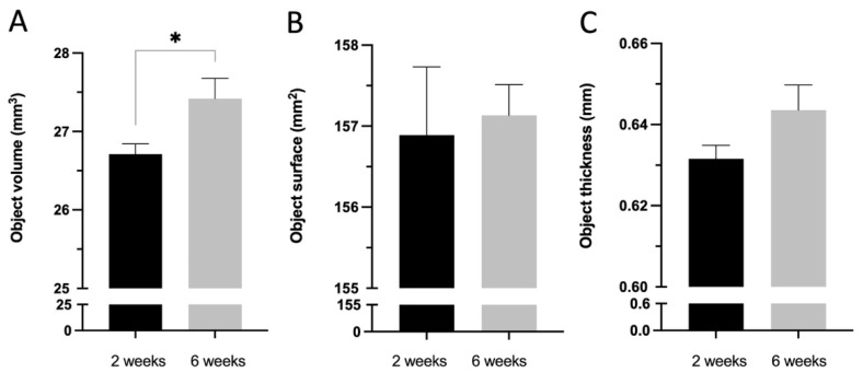
Suture anchors are extensively used in rotator cuff tear surgery. With the advancement of three-dimensional printing technology, biodegradable metal has been developed for orthopedic applications. This study adopted three-dimensional-printed biodegradable Fe suture anchors with double-helical threads and commercialized non-vented screw-type Ti suture anchors with a tapered tip in the experimental and control groups, respectively. The in vitro study showed that the Fe and Ti suture anchors exhibited a similar ultimate failure load in 20-pound-per-cubic-foot polyurethane foam blocks and rabbit bone. In static immersion tests, the corrosion rate of Fe suture anchors was 0.049 ± 0.002 mm/year. The in vivo study was performed on New Zealand white rabbits and SAs were employed to reattach the ruptured supraspinatus tendon. The in vivo ultimate failure load of the Fe suture anchors was superior to that of the Ti suture anchors at 6 weeks. Micro-computed tomography showed that the bone volume fraction and bone surface density in the Fe suture anchors group 2 and 6 weeks after surgery were superior, and the histology confirmed that the increased bone volume around the anchor was attributable to mineralized osteocytes. The three-dimensional-printed Fe suture anchors outperformed the currently used Ti suture anchors.
缝线锚钉在肩袖撕裂手术中被广泛应用。随着三维打印技术的发展,可生物降解金属已被开发用于骨科应用。本研究在实验组和对照组分别采用了具有双螺旋螺纹的三维打印可生物降解铁缝线锚钉和商业化的带锥形尖端的非通风螺旋型钛缝线锚钉。体外研究表明,铁和钛缝线锚钉在每立方英尺20磅的聚氨酯泡沫块和兔骨中表现出相似的极限破坏载荷。在静态浸泡试验中,铁缝线锚钉的腐蚀速率为0.049±0.002毫米/年。体内研究在新西兰白兔身上进行,使用缝线锚钉重新附着断裂的冈上肌腱。铁缝线锚钉在6周时的体内极限破坏载荷优于钛缝线锚钉。微型计算机断层扫描显示,术后2周和6周时铁缝线锚钉组的骨体积分数和骨表面密度更高,组织学证实锚钉周围骨体积的增加归因于矿化的骨细胞。三维打印的铁缝线锚钉性能优于目前使用的钛缝线锚钉。