Lambova Sevdalina Nikolova, Kurteva Ekaterina Krasimirova, Dzhambazova Sanie Syuleymanova, Vasilev Georgi Hristov, Kyurkchiev Dobroslav Stanimirov, Geneva-Popova Mariela Gencheva
Department of Propaedeutics of Internal Diseases, Faculty of Medicine, Medical University of Plovdiv, 4002 Plovdiv, Bulgaria.
Department of Rheumatology, MHAT "Sveti Mina", 4000 Plovdiv, Bulgaria.
Life (Basel). 2022 Mar 29;12(4):498. doi: 10.3390/life12040498.
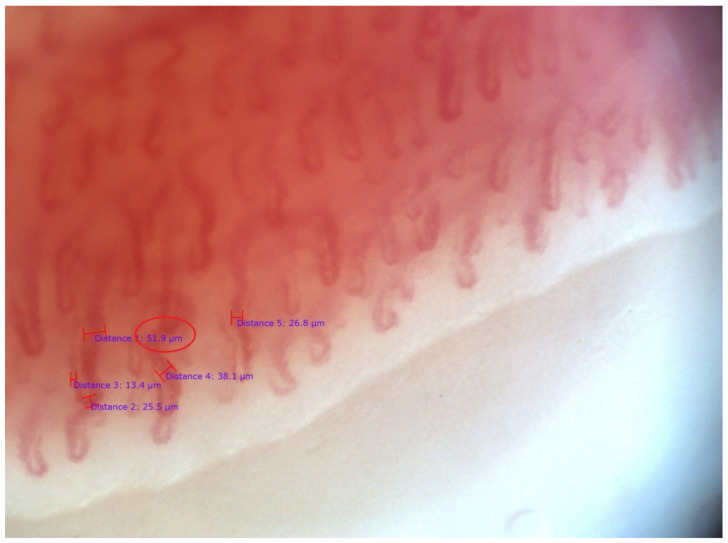
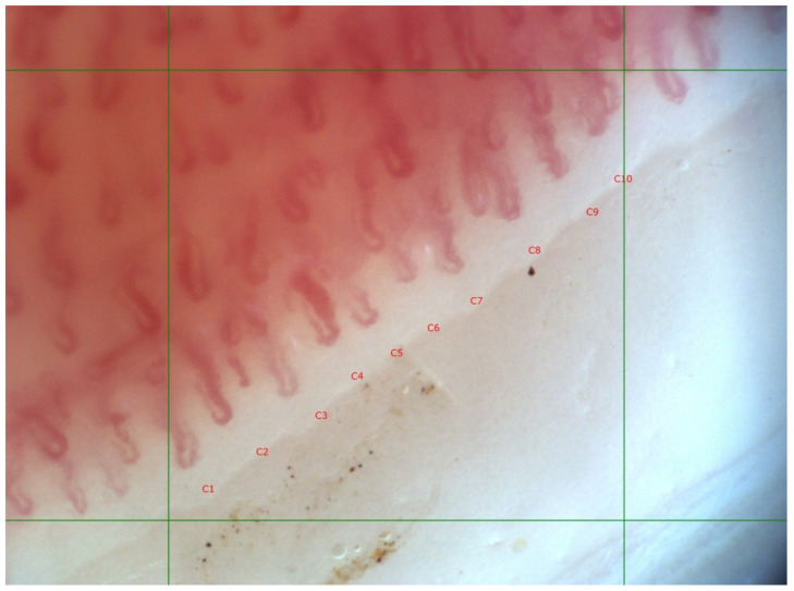
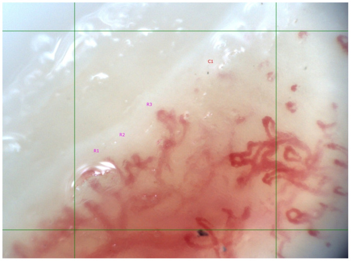
Introduction: Data on the associations between capillaroscopic changes and diagnostic systemic-sclerosis (SSc)-related antibodies are scarce. Presence of such correlation would improve current knowledge about the disease’s pathogenesis by revealing the mechanisms of microangiopathy. The microvascular pathology of SSc is a hallmark of the disease, and immunological abnormalities probably contribute to its development. Patients and methods: 19 patients with definite diagnosis of SSc were included in the current pilot study; 16 had limited and 3 had diffuse cutaneous involvement; their mean age was 51.56 ± 15.07 years. All patients exhibited symptoms of Raynaud’s phenomenon of the fingers. A “scleroderma” type capillaroscopic pattern was classified according to the staging suggested by Cutolo et al. (2000): “early”, “active” or ”late” phase. In the presence of different degrees of capillaroscopic changes in different fingers, the most-advanced microvascular pathology was chosen for classification. In cases without capillaroscopic features of microangiopathy, the findings were categorized as normal or nonspecific (dilated, tortuous capillaries, and/or hemorrhages). Indirect immunofluorescence on HEp-2 cells was performed as the gold-standard screening method for the detection of antinuclear autoantibodies (ANA), and determination of the immunofluorescent staining pattern (anti-cell pattern) was in accordance with the International Consensus on ANA Patterns. Scleroderma-associated autoantibodies in the patients’ serum were assessed using line immunoblot assay for detection of autoantibodies to 13 scleroderma-associated autoantigens: Scl-70, CENP A, CENP B, RP11/RNAP-III, RP155/RNAP-III, fibrillarin, NOR-90, Th/To, PM-Scl100, PM-Scl75, Ku, PDGFR, and Ro-52. Results: In 73.7% (n = 14) of the examined patients, “scleroderma” type capillaroscopic changes were found, and in 26.3% (n = 5), capillaroscopic features of microangiopathy were absent (nonspecific changes, n = 3; normal findings, n = 2). In SSc patients with positive anti-Scl-70 (n = 7) antibodies, significantly lower mean capillary density was observed along with a higher frequency of “active” and “late” phase capillaroscopic changes as compared to the anti-Scl-70-negative patients (p < 0.05). Anti-RNAP III−155 positive patients (n = 4) had significantly higher mean capillary density than anti-RNAP III−155 negative patients (n = 15). In three of the anti-RNAP III−155-positive cases, capillaroscopic features of microangiopathy were not detected, and in one case there was an “early” phase “scleroderma” pattern. Conclusion: In the current pilot study, the association between more advanced capillaroscopic changes and the presence of anti-Scl-70 autoantibodies was confirmed. As a novel observation, positive anti-RNAP III−155 antibodies were found in SSc patients with or without early microangiopathy. The question of associations between microvascular changes in SSc and other SSc-related autoantibodies requires further research.
关于毛细血管镜检查变化与诊断系统性硬化症(SSc)相关抗体之间关联的数据很少。这种相关性的存在将通过揭示微血管病变的机制来增进我们对该疾病发病机制的现有认识。SSc的微血管病理是该疾病的一个标志,免疫异常可能在其发展过程中起作用。
本初步研究纳入了19例确诊为SSc的患者;16例为局限性皮肤受累,3例为弥漫性皮肤受累;他们的平均年龄为51.56±15.07岁。所有患者均表现出手指雷诺现象的症状。根据Cutolo等人(2000年)建议的分期,将“硬皮病”型毛细血管镜检查模式分为“早期”、“活动期”或“晚期”阶段。在不同手指存在不同程度的毛细血管镜检查变化时,选择最严重的微血管病理进行分类。在无微血管病变毛细血管镜特征的病例中,检查结果分类为正常或非特异性(扩张、迂曲的毛细血管和/或出血)。采用间接免疫荧光法检测HEp-2细胞上的抗核自身抗体(ANA),作为检测抗核抗体的金标准筛查方法,并根据抗核抗体模式国际共识确定免疫荧光染色模式(抗细胞模式)。使用线性免疫印迹法检测患者血清中的硬皮病相关自身抗体,以检测针对13种硬皮病相关自身抗原的自身抗体:Scl-70、CENP A、CENP B、RP11/RNAP-III、RP155/RNAP-III、原纤维蛋白、NOR-90、Th/To、PM-Scl100、PM-Scl75、Ku、PDGFR和Ro-52。
在73.7%(n = 14)的受检患者中发现了“硬皮病”型毛细血管镜检查变化,26.3%(n = 5)的患者无微血管病变的毛细血管镜特征(非特异性变化,n = 3;正常结果,n = 2)。与抗Scl-70抗体阴性的患者相比,抗Scl-70抗体阳性(n = 7)的SSc患者平均毛细血管密度显著降低,同时“活动期”和“晚期”毛细血管镜检查变化的频率更高(p < 0.05)。抗RNAP III-155阳性患者(n = 4)的平均毛细血管密度显著高于抗RNAP III-155阴性患者(n = 15)。在3例抗RNAP III-1