Fava Andrea, Cimbro Raffaello, Wigley Fredrick M, Liu Qing-Rong, Rosen Antony, Boin Francesco
Department of Medicine, Division of Rheumatology, Johns Hopkins University School of Medicine, 5200 Eastern Avenue, MFL Building, Center Tower, Suite 4100, Baltimore, MD, 21224, USA.
Behavioral Neuroscience Research Branch, National Institute of Drug Abuse, National Institutes of Health, 251 Bayview Boulevard, Baltimore, Maryland, 21224, USA.
Arthritis Res Ther. 2016 May 4;18(1):99. doi: 10.1186/s13075-016-0993-2.
Scleroderma is an antigen-driven T cell-mediated autoimmune disease. Presence of anti-topoisomerase-I antibodies is associated with pulmonary fibrosis and predicts increased mortality. Characterization of autoreactive T lymphocytes may shed light on disease pathogenesis and serve as a biomarker for disease activity. Here, we aimed to quantify and functionally characterize circulating topoisomerase I (topo-I)-specific CD4+ T cells and to define their association with presence and progression of interstitial lung disease (ILD) in patients with scleroderma.
Using flow cytometry, circulating topo-I-reactive CD4+ T cells were identified by the expression of specific activation markers (CD154 and CD69) upon stimulation with purified topo-I and quantified in 27 SSc patients and 4 healthy donors (HD). Polarization of autoreactive T cells (Th1, Th2, Th17, Th1-17) was defined using surface expression of specific chemokine receptors. Presence and progression of ILD were determined using high-resolution chest CT and pulmonary function tests.
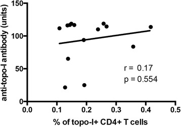
Topo-I-reactive CD4+ T cells were found in all topo-I-positive patients compared to one topo-I-negative subject and no HD. Topo-I-specific CD4+ T cells exhibited a distinct Th17 polarized phenotype. Autoreactive T cells were significantly increased in subjects with evidence of ILD and were quantitatively associated with the decline of lung volumes.
Topo-I-specific T cells can be reliably quantified in the peripheral blood of patients with scleroderma, exhibit a pro-inflammatory Th17 phenotype, and predict progression of ILD.
硬皮病是一种抗原驱动的T细胞介导的自身免疫性疾病。抗拓扑异构酶-I抗体的存在与肺纤维化相关,并预示死亡率增加。自身反应性T淋巴细胞的特征可能有助于揭示疾病发病机制,并作为疾病活动的生物标志物。在此,我们旨在量化并从功能上表征循环中的拓扑异构酶I(topo-I)特异性CD4 + T细胞,并确定它们与硬皮病患者间质性肺疾病(ILD)的存在和进展之间的关联。
使用流式细胞术,通过用纯化的topo-I刺激后特定激活标志物(CD154和CD69)的表达来鉴定循环中的topo-I反应性CD4 + T细胞,并在27例系统性硬化症(SSc)患者和4名健康供体(HD)中进行量化。使用特定趋化因子受体的表面表达来定义自身反应性T细胞(Th1、Th2、Th17、Th1-17)的极化。使用高分辨率胸部CT和肺功能测试来确定ILD的存在和进展。
与一名topo-I阴性受试者和无HD相比,在所有topo-I阳性患者中均发现了topo-I反应性CD4 + T细胞。topo-I特异性CD4 + T细胞表现出独特的Th17极化表型。在有ILD证据的受试者中,自身反应性T细胞显著增加,并且在数量上与肺容积的下降相关。
在硬皮病患者的外周血中可以可靠地量化topo-I特异性T细胞,其表现出促炎性Th17表型,并可预测ILD的进展。