Lever Mael, Chen Ying, Glaser Moritz, Unterlauft Jan Darius, Lommatzsch Claudia, Bechrakis Nikolaos E, Böhm Michael R R
Department of Ophthalmology, University Hospital Essen, 45147 Essen, Germany.
Achim Wessing Institute for Imaging in Ophthalmology, University Hospital Essen, 45147 Essen, Germany.
Life (Basel). 2022 Apr 11;12(4):568. doi: 10.3390/life12040568.
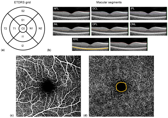
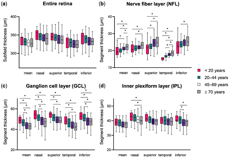
In ocular, neurologic, and cardiovascular diseases, macular segmentation data from spectral-domain optical coherence tomography (SD-OCT) provide morphologic, and OCT-angiography (OCTA) results give microvascular information about the macula. Age was shown to influence both methods' measurements. To further characterize this association, macular SD-OCT and OCTA changes were investigated in a population of juvenile, adult, and older individuals. Macular segment thickness and superficial (SCP) and deep plexus (DCP) vascular density (VD) of 157 healthy individuals aged 10-79 years were analyzed retrospectively. One-way analysis of variance (ANOVA) was used to compare age groups. The association between macular segmentation and OCTA parameters and between these and age was evaluated using linear regression. ANOVA and linear regression analysis showed a thickness decrease in the whole macular and in the ganglion cell and inner plexiform layers with age. While the foveal avascular zone area remained constant between age groups, VD of the SCP and DCP also decreased with age. In multiple linear regression, SCP and DCP VD were associated with inner macular segment thickness in an age-independent way. To conclude, the age-related microvascular and morphological changes in the macula described in this study can contribute to improving the understanding of macular aging processes and better interpreting OCT(A) results in healthy individuals and patients suffering from various retinal diseases.
在眼部、神经和心血管疾病中,光谱域光学相干断层扫描(SD-OCT)的黄斑分割数据提供形态学信息,而光学相干断层扫描血管造影(OCTA)结果则给出黄斑的微血管信息。研究表明年龄会影响这两种方法的测量结果。为了进一步描述这种关联,我们在青少年、成年人和老年人群体中研究了黄斑SD-OCT和OCTA的变化。回顾性分析了157名年龄在10 - 79岁的健康个体的黄斑段厚度、浅层(SCP)和深层丛状层(DCP)血管密度(VD)。采用单因素方差分析(ANOVA)比较年龄组。使用线性回归评估黄斑分割与OCTA参数之间以及这些参数与年龄之间的关联。方差分析和线性回归分析表明,随着年龄增长,整个黄斑以及神经节细胞层和内丛状层的厚度会降低。虽然年龄组之间的中心凹无血管区面积保持不变,但SCP和DCP的VD也随年龄降低。在多元线性回归中,SCP和DCP的VD与黄斑内段厚度以年龄无关的方式相关。总之,本研究中描述的黄斑与年龄相关的微血管和形态学变化有助于增进对黄斑老化过程的理解,并更好地解释健康个体和患有各种视网膜疾病患者的OCT(A)结果。