Beijing Tongren Eye Center, Beijing Tongren Hospital, Beijing Ophthalmology and Visual Science Key Lab, Beijing Key Laboratory of Intraocular Tumor Diagnosis and Treatment, Capital Medical University, 1 Dong Jiao Min Xiang, Dong Cheng District, Beijing, 100730, China.
Beijing Institute of Ophthalmology, Beijing Ophthalmology and Visual Science Key Lab, Beijing Tongren Eye Center, Beijing Tongren Hospital, Capital Medical University, 17 Hougou Lane, Chong Wen Men, Beijing, 100005, China.
BMC Ophthalmol. 2020 Feb 12;20(1):49. doi: 10.1186/s12886-019-1296-6.
Diagnosis and follow-up of retinal diseases may be improved if the thickness of the various retinal layers, in addition to the total retinal thickness, is taken into account. Here we measured the thickness of the macular retinal layers in a population-based study group to assess the normative values and their associations.
Using spectral-domain optical coherence tomographic images (Spectralis®, wavelength: 870 nm; Heidelberg Engineering Co, Heidelberg, Germany), we measured the thickness of the macular retinal layers in participants of the population-based Beijing Eye Study without ocular diseases and without systematic diseases, such as arterial hypertension, hyperlipidemia, diabetes mellitus, cardiovascular diseases, previous myocardial infarction, cerebral trauma and stroke. Segmentation and measurement of the retinal layers was performed automatically in each of the horizontal scans.
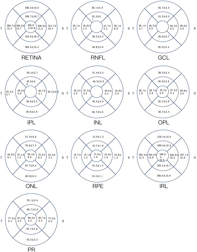
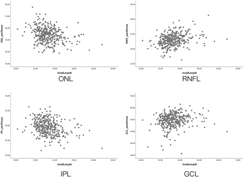
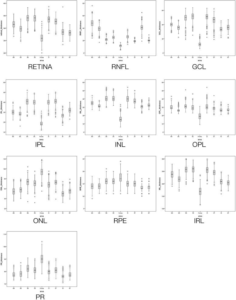
The study included 384 subjects (mean age:60.0 ± 8.0 years). The mean thickness of the whole retina, outer plexiform layer, outer nuclear layer,retinal pigment epithelium, inner retinal layer and photoreceptor layer was 259.8 ± 18.9 μm, 19.4 ± 3.9 μm, 93.4 ± 9.6 μm, 17.6 ± 1.9 μm, 169.8 ± 18.6 μm, and 90.0 ± 4.2 μm, respectively. In multivariable analysis, the thickness of the foveola and of all retinal layers in the foveal, parafoveal and perifoveal region decreased with older age (all P < 0.05), except for the thickness of the parafoveal outer plexiform layer which increased with age. Men as compared to women had higher thickness measurements of the photoreceptor layer and outer nuclear layer in all areas, and of all layers between the retinal nerve fiber layer and inner nuclear layer in the parafoveal area (all P < 0.05). The associations between the macular retinal layers thickness and axial length were not consistent. The inner plexiform layer was thicker, and the ganglion cell layer and inner nuclear layer were thinner, in the temporal areas than in the nasal areas, CONCLUSIONS: The associations between decreasing thickness of most retinal layers with older age and the correlation of a higher thickness of some retinal layers with male gender may clinically be taken into account.
除了总视网膜厚度外,如果能考虑到各视网膜层的厚度,那么对视网膜疾病的诊断和随访可能会得到改善。在此,我们在一个基于人群的研究组中测量了黄斑视网膜层的厚度,以评估其正常值及其相关性。
使用频域光学相干断层扫描图像(Spectralis®, 波长:870nm;Heidelberg Engineering Co,Heidelberg,德国),我们在没有眼部疾病和系统性疾病(如动脉高血压、高血脂、糖尿病、心血管疾病、既往心肌梗死、脑外伤和中风)的基于人群的北京眼研究参与者中测量了黄斑视网膜层的厚度。在每个水平扫描中自动对视网膜层进行分割和测量。
该研究纳入了 384 名受试者(平均年龄:60.0±8.0 岁)。整个视网膜、外丛状层、外核层、视网膜色素上皮、内视网膜层和光感受器层的平均厚度分别为 259.8±18.9μm、19.4±3.9μm、93.4±9.6μm、17.6±1.9μm、169.8±18.6μm 和 90.0±4.2μm。在多变量分析中,除了黄斑区外丛状层厚度随年龄增长而增加外,黄斑区和旁黄斑区及中心凹周围视网膜层的厚度均随年龄增长而降低(均 P<0.05)。与女性相比,男性在所有区域的光感受器层和外核层以及旁黄斑区神经纤维层和内核层之间的所有层的厚度测量值均较高(均 P<0.05)。黄斑区视网膜层厚度与眼轴的相关性并不一致。颞侧区域的内丛状层较厚,神经节细胞层和内核层较薄。
随着年龄的增长,大多数视网膜层厚度降低,以及某些视网膜层厚度与男性相关,这些相关性可能会在临床上得到考虑。