Department of Ophthalmology, The Second Hospital of Jilin University, Changchun, China.
BMC Ophthalmol. 2022 Apr 22;22(1):184. doi: 10.1186/s12886-022-02355-5.
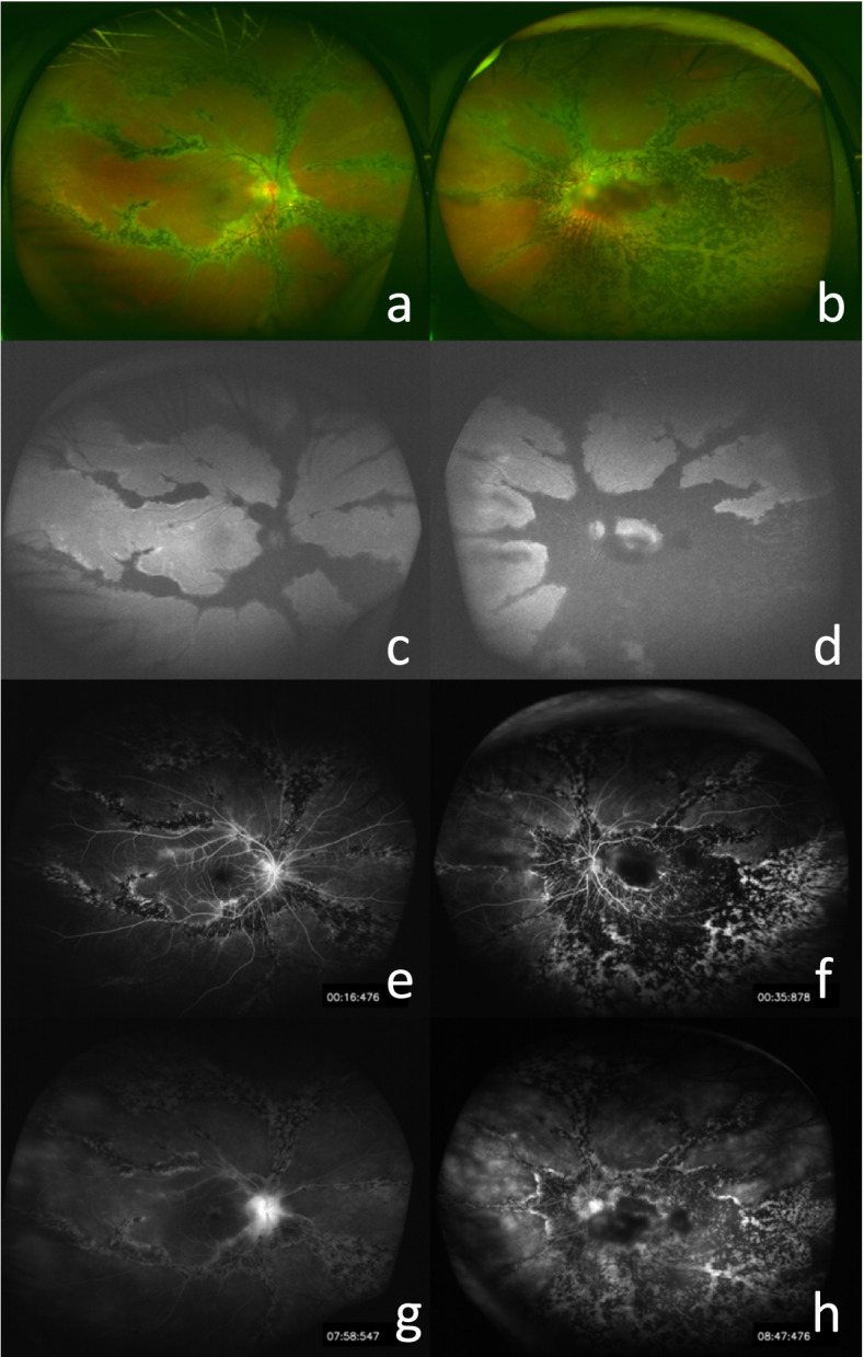
Pigmented paravenous retinochoroidal atrophy (PPRCA) is a rare fundus disease characterized by the presence of osteoblast-like pigment, atrophy of retinal pigment epithelium (RPE), and choroid deposition along the large retinal veins.
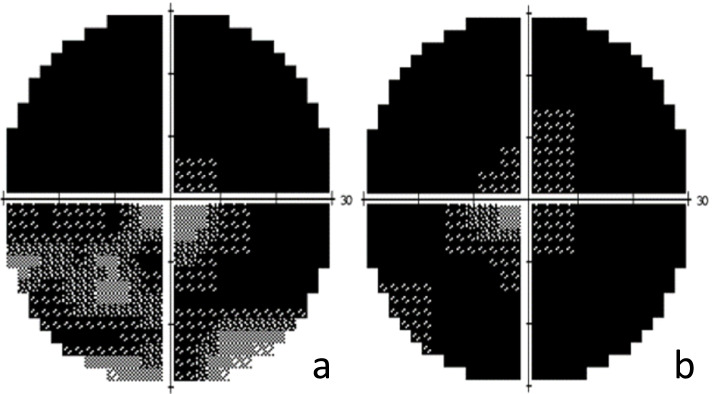
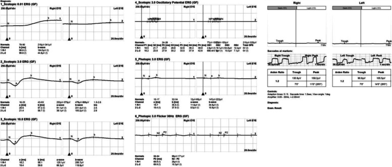
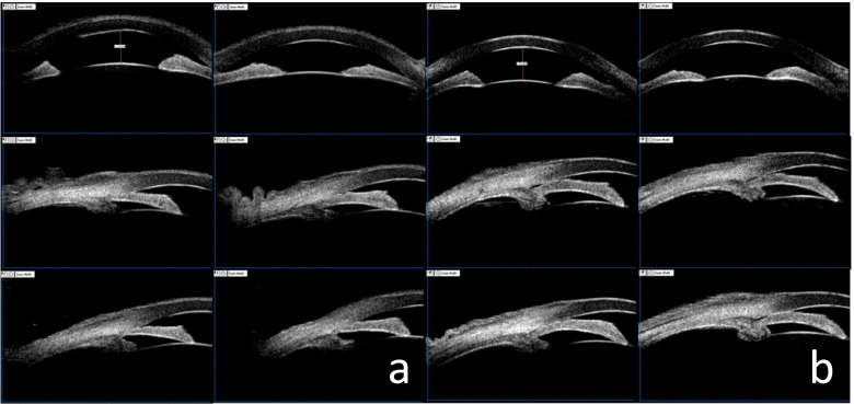
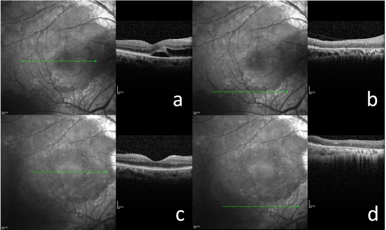
A 55-year-old Chinese female presented with right eye distention and bilateral vision loss. Osteocyte-like pigmentation and retinal choroidal atrophy distributed along the large retinal veins were seen in the fundus of bilateral eyes. The atrophy in the left eye was more severe compared to the right eye. The patient also presented with bilateral acute angle-closure glaucoma (AACG) and posterior subcapsular cataract (PSC) accompanied with anterior segmental manifestations, similar to the complications of retinitis pigmentosa (RP). The patient underwent ultrasound biomicroscopy (UBM), Humphrey field analyser (HFA), optical coherence tomography (OCT), fundus autofluorescence (FAF), fluorescein fundus angiography (FFA), electroretinogram (ERG), and electrooculography (EOG), all of which confirmed the aforementioned diagnose.
PPRCA is a rare disease of unknown etiology. The patient in this case presented with complications similar to those of RP, and the two conditions may share a genetic basis. Further studies are needed to confirm this relationship.
色素性静脉旁视网膜脉络膜萎缩(PPRCA)是一种罕见的眼底疾病,其特征为存在成骨样色素、视网膜色素上皮(RPE)萎缩以及脉络膜沿大的视网膜静脉沉积。
一名 55 岁的中国女性因右眼突出和双眼视力下降就诊。双眼眼底可见沿大的视网膜静脉分布的骨细胞样色素沉着和视网膜脉络膜萎缩。左眼的萎缩比右眼更严重。患者还患有双侧急性闭角型青光眼(AACG)和后发性白内障(PSC),伴有前段表现,类似于色素性视网膜炎(RP)的并发症。患者接受了超声生物显微镜(UBM)、Humphrey 视野分析仪(HFA)、光学相干断层扫描(OCT)、眼底自发荧光(FAF)、荧光素眼底血管造影(FFA)、视网膜电图(ERG)和眼电图(EOG)检查,所有检查均证实了上述诊断。
PPRCA 是一种病因不明的罕见疾病。本例患者表现出类似于 RP 的并发症,这两种疾病可能具有遗传基础。需要进一步的研究来证实这种关系。