• 文献检索
  • 文档翻译
  • 深度研究
  • 学术资讯
  • Suppr Zotero 插件Zotero 插件
  • 邀请有礼
  • 套餐&价格
  • 历史记录
应用&插件
Suppr Zotero 插件Zotero 插件浏览器插件Mac 客户端Windows 客户端微信小程序
定价
高级版会员购买积分包购买API积分包
服务
文献检索文档翻译深度研究API 文档MCP 服务
关于我们
关于 Suppr公司介绍联系我们用户协议隐私条款
关注我们

Suppr 超能文献

核心技术专利:CN118964589B侵权必究
粤ICP备2023148730 号-1Suppr @ 2026

文献检索

告别复杂PubMed语法,用中文像聊天一样搜索,搜遍4000万医学文献。AI智能推荐,让科研检索更轻松。

立即免费搜索

文件翻译

保留排版,准确专业,支持PDF/Word/PPT等文件格式,支持 12+语言互译。

免费翻译文档

深度研究

AI帮你快速写综述,25分钟生成高质量综述,智能提取关键信息,辅助科研写作。

立即免费体验

色素性静脉旁脉络膜视网膜萎缩:最新情况

Pigmented paravenous chorioretinal atrophy: Updated scenario.

作者信息

Antropoli Alessio, Arrigo Alessandro, Pili Lorenzo, Bianco Lorenzo, Berni Alessandro, Saladino Andrea, Bandello Francesco, Battaglia Parodi Maurizio

机构信息

Department of Ophthalmology, IRCCS San Raffaele Scientific Institute, Milan, Italy.

出版信息

Eur J Ophthalmol. 2024 Jul;34(4):941-951. doi: 10.1177/11206721231199118. Epub 2023 Sep 5.

DOI:10.1177/11206721231199118
PMID:37670517
原文链接:https://pmc.ncbi.nlm.nih.gov/articles/PMC11295417/
Abstract

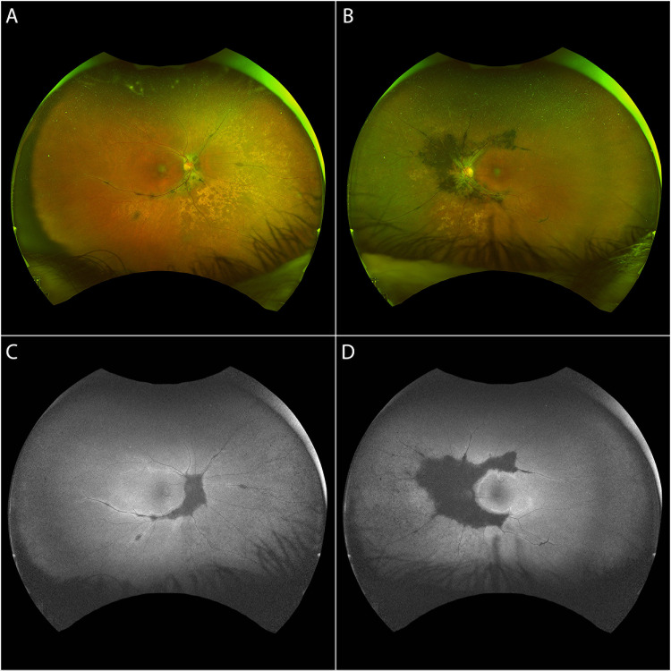
Pigmented paravenous chorioretinal atrophy (PPCRA) is an uncommon form of chorioretinal atrophy characterized by perivenous aggregations of pigment clumps associated with peripapillary and radial zones of retinal pigment epithelial atrophy that are distributed along the retinal veins. Most patients are asymptomatic, and evidence suggest that PPCRA is slowly progressing. Unless macular involvement is present, the majority of patients usually retain a normal visual function. Our ability to diagnose PPCRA has recently improved thanks to multimodal imaging, especially with the advent of ultra-widefield (UWF) imaging. Blood tests and functional and genetic testing can help with the correct differential diagnosis of pseudo-PPCRA or other disorders with similar characteristics. Although the cause of PPCRA is unknown, it is possible that it has a genetic basis. In this review we provide a summary of the multimodal imaging characteristics of PPCRA, and discuss its possible pathogenesis, based on the genes that have been associated with this disease.

摘要

色素性静脉旁脉络膜视网膜萎缩(PPCRA)是一种罕见的脉络膜视网膜萎缩形式,其特征是色素团块沿视网膜静脉呈静脉周围聚集,并伴有视乳头周围和视网膜色素上皮萎缩的放射状区域。大多数患者无症状,有证据表明PPCRA进展缓慢。除非累及黄斑,大多数患者通常保留正常视觉功能。由于多模态成像,特别是超广域(UWF)成像的出现,我们诊断PPCRA的能力最近有所提高。血液检查以及功能和基因检测有助于对假性PPCRA或其他具有相似特征的疾病进行正确的鉴别诊断。虽然PPCRA的病因尚不清楚,但有可能具有遗传基础。在本综述中,我们总结了PPCRA的多模态成像特征,并根据与该疾病相关的基因讨论其可能的发病机制。

https://cdn.ncbi.nlm.nih.gov/pmc/blobs/1036/11295417/2e5d764a92c5/10.1177_11206721231199118-fig3.jpg
https://cdn.ncbi.nlm.nih.gov/pmc/blobs/1036/11295417/d9acbbb69989/10.1177_11206721231199118-fig1.jpg
https://cdn.ncbi.nlm.nih.gov/pmc/blobs/1036/11295417/dd59239c1fd1/10.1177_11206721231199118-fig2.jpg
https://cdn.ncbi.nlm.nih.gov/pmc/blobs/1036/11295417/2e5d764a92c5/10.1177_11206721231199118-fig3.jpg

相似文献

1
Pigmented paravenous chorioretinal atrophy: Updated scenario.色素性静脉旁脉络膜视网膜萎缩:最新情况
Eur J Ophthalmol. 2024 Jul;34(4):941-951. doi: 10.1177/11206721231199118. Epub 2023 Sep 5.
2
Pigmented paravenous chorioretinal atrophy with Coat's like response.色素性脉络膜视网膜旁萎缩伴 Coat 样反应。
Indian J Ophthalmol. 2013 Oct;61(10):586-8. doi: 10.4103/0301-4738.121083.
3
Pigmented Paravenous Chorioretinal Atrophy: Clinical Spectrum and Multimodal Imaging Characteristics.色素性静脉旁脉络膜视网膜萎缩:临床谱及多模态影像学特征
Am J Ophthalmol. 2021 Apr;224:120-132. doi: 10.1016/j.ajo.2020.12.010. Epub 2020 Dec 16.
4
Pigmented Paravenous Chorioretinal Atrophy (PPCRA).色素性静脉旁视网膜脉络膜萎缩(PPCRA)。
Adv Exp Med Biol. 2018;1085:111-113. doi: 10.1007/978-3-319-95046-4_22.
5
Pigmented paravenous retinochoroidal atrophy in an infant with unilateral macular involvement: case report and literature review.一名单侧黄斑受累婴儿的色素性静脉旁视网膜脉络膜萎缩:病例报告及文献综述
Can J Ophthalmol. 2024 Jun;59(3):e287-e289. doi: 10.1016/j.jcjo.2023.11.018. Epub 2023 Dec 16.
6
Fundus autofluorescence and optical coherence tomography findings in pigmented paravenous retinochoroidal atrophy.色素性静脉旁视网膜脉络膜萎缩的眼底自发荧光和光学相干断层扫描结果
Can J Ophthalmol. 2014 Dec;49(6):e144-6. doi: 10.1016/j.jcjo.2014.08.019.
7
PERIPHERAL RETINAL VASCULAR ABNORMALITIES IN PIGMENTED PARAVENOUS RETINOCHOROIDAL ATROPHY.色素性静脉旁视网膜脉络膜萎缩的周边视网膜血管异常
Am J Ophthalmol. 2022 Apr;236:e4-e5. doi: 10.1016/j.ajo.2022.01.003. Epub 2022 Jan 14.
8
[Pigmented paravenous retinochoroidal atrophy--a case report].[色素性静脉旁视网膜脉络膜萎缩——病例报告]
Klin Oczna. 2014;116(2):115-8.
9
Pigmented paravenous chorioretinal atrophy is associated with a mutation within the crumbs homolog 1 (CRB1) gene.色素性静脉旁脉络膜视网膜萎缩与crumbs同源物1(CRB1)基因的突变有关。
Invest Ophthalmol Vis Sci. 2005 Jan;46(1):322-8. doi: 10.1167/iovs.04-0734.
10
Pigmented paravenous retinochoroidal atrophy: a case report.色素性静脉旁视网膜脉络膜萎缩:一例报告
BMC Ophthalmol. 2018 Jun 7;18(1):136. doi: 10.1186/s12886-018-0809-z.

引用本文的文献

1
Bilateral Perivascular Chorioretinal Atrophy Resembling Pigmented Paravenous Chorioretinal Atrophy Post COVID-19 Infection: A Case Report and Comprehensive Immune Profiling.新冠病毒感染后双侧血管周围脉络膜视网膜萎缩,类似色素性静脉旁脉络膜视网膜萎缩:一例报告及全面免疫分析
Vaccines (Basel). 2024 Aug 2;12(8):878. doi: 10.3390/vaccines12080878.
2
Relationship between ischemic index, leakage index, and macular edema in branch retinal vein occlusion.分支型视网膜静脉阻塞中缺血指数、渗漏指数与黄斑水肿的关系。
Graefes Arch Clin Exp Ophthalmol. 2024 May;262(5):1499-1506. doi: 10.1007/s00417-023-06343-3. Epub 2023 Dec 26.
3

本文引用的文献

1
Posterior vitreous cortex hyalocytes visualization in asymmetric pigmented paravenous chorioretinal atrophy (PPCRA) using OCT.使用光学相干断层扫描(OCT)观察不对称性色素性静脉旁脉络膜视网膜萎缩(PPCRA)中的玻璃体后皮质玻璃体细胞。
Am J Ophthalmol Case Rep. 2023 Apr 12;30:101846. doi: 10.1016/j.ajoc.2023.101846. eCollection 2023 Jun.
2
variant associated with pigmented paravenous chorioretinal atrophy.与色素性静脉旁脉络膜视网膜萎缩相关的变体
Eur J Ophthalmol. 2023 Nov;33(6):NP6-NP9. doi: 10.1177/11206721231155042. Epub 2023 Feb 8.
3
Identifying two pathogenic variants in a patient with pigmented paravenous retinochoroidal atrophy.
Association of Circulating Antiretinal Antibodies With Clinical Outcomes in Retinitis Pigmentosa.
循环抗视网膜抗体与色素性视网膜炎临床结局的关联。
Invest Ophthalmol Vis Sci. 2023 Dec 1;64(15):13. doi: 10.1167/iovs.64.15.13.
4
A Case of Pigmented Paravenous Retinochoroidal Atrophy With Retinitis Pigmentosa.一例伴有色素性视网膜炎的色素性静脉旁视网膜脉络膜萎缩病例。
Cureus. 2023 Nov 8;15(11):e48532. doi: 10.7759/cureus.48532. eCollection 2023 Nov.
在一名患有色素性静脉旁视网膜脉络膜萎缩的患者中鉴定出两个致病变体。
Open Life Sci. 2023 Jan 16;18(1):20220532. doi: 10.1515/biol-2022-0532. eCollection 2023.
4
Pigmented Paravenous Chorioretinal Atrophy With Macular Hole.伴有黄斑裂孔的色素性静脉旁脉络膜视网膜萎缩
JAMA Ophthalmol. 2022 Aug 1;140(8):e222437. doi: 10.1001/jamaophthalmol.2022.2437. Epub 2022 Aug 18.
5
Identification of Arhgef12 and Prkci as genetic modifiers of retinal dysplasia in the Crb1rd8 mouse model.鉴定 Arhgef12 和 Prkci 作为 Crb1rd8 小鼠模型视网膜发育不良的遗传修饰因子。
PLoS Genet. 2022 Jun 8;18(6):e1009798. doi: 10.1371/journal.pgen.1009798. eCollection 2022 Jun.
6
Pigmented paravenous retinochoroidal atrophy with acute angle-closure glaucoma and posterior subcapsular cataract: a case report.色素性静脉旁视网膜脉络膜萎缩伴急性闭角型青光眼和后囊下白内障:一例报告。
BMC Ophthalmol. 2022 Apr 22;22(1):184. doi: 10.1186/s12886-022-02355-5.
7
PERIPHERAL RETINAL VASCULAR ABNORMALITIES IN PIGMENTED PARAVENOUS RETINOCHOROIDAL ATROPHY.色素性静脉旁视网膜脉络膜萎缩的周边视网膜血管异常
Am J Ophthalmol. 2022 Apr;236:e4-e5. doi: 10.1016/j.ajo.2022.01.003. Epub 2022 Jan 14.
8
OPTICAL COHERENCE TOMOGRAPHY ANGIOGRAPHY FINDINGS IN PIGMENTED PARAVENOUS CHORIORETINAL ATROPHY.光学相干断层扫描血管造影在色素性脉络膜视网膜萎缩中的发现。
Retina. 2022 May 1;42(5):915-922. doi: 10.1097/IAE.0000000000003407.
9
Pigmented Paravenous Retinochoroidal Atrophy: A Case Report Supported by Multimodal Imaging Studies.色素性脉络膜视网膜萎缩:多模态影像学研究支持的一例报告。
Medicina (Kaunas). 2021 Dec 19;57(12):1382. doi: 10.3390/medicina57121382.
10
Fundus autofluorescence detects subtle pigmentary alterations in pigmented paravenous retinochoroidal atrophy.眼底自发荧光可检测色素性静脉旁视网膜脉络膜萎缩中的细微色素改变。
Am J Ophthalmol Case Rep. 2021 Jul 3;23:101158. doi: 10.1016/j.ajoc.2021.101158. eCollection 2021 Sep.