Palamà Ilaria Elena, Maiorano Gabriele, Di Maria Francesca, Zangoli Mattia, Candini Andrea, Zanelli Alberto, D'Amone Stefania, Fabiano Eduardo, Gigli Giuseppe, Barbarella Giovanna
Nanotechnology Institute (CNR-NANOTEC) and Department of Mathematics and Physics, University of Salento, Monteroni Street, 73100 Lecce, Italy.
CNR-ISOF and Mediteknology srl Area Ricerca CNR, Piero Gobetti Street 101, 40129 Bologna, Italy.
ACS Omega. 2022 Apr 6;7(15):12624-12636. doi: 10.1021/acsomega.1c06677. eCollection 2022 Apr 19.
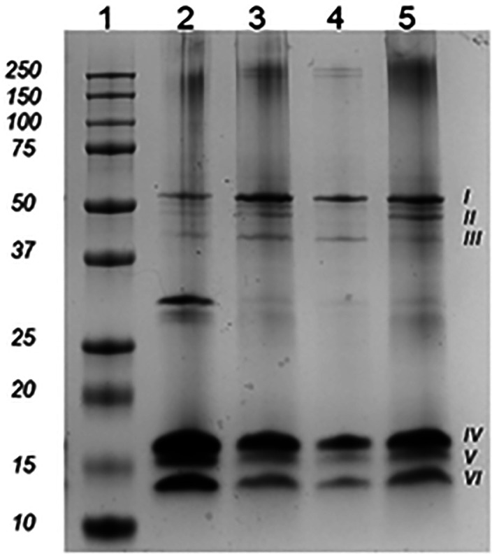
Protein-based microfibers are biomaterials of paramount importance in materials science, nanotechnology, and medicine. Here we describe the spontaneous in situ formation and secretion of nanostructured protein microfibers in 2D and 3D cell cultures of 3T3 fibroblasts and B104 neuroblastoma cells upon treatment with a micromolar solution of either unmodified terthiophene or terthiophene modified by mono-oxygenation (thiophene → thiophene -oxide) or dioxygenation (thiophene → thiophene ,-dioxide) of the inner ring. We demonstrate via metabolic cytotoxicity tests that modification to the -oxide leads to a severe drop in cell viability. By contrast, unmodified terthiophene and the respective ,-dioxide cause no harm to the cells and lead to the formation and secretion of fluorescent and electroactive protein-fluorophore coassembled microfibers with a large aspect ratio, a micrometer-sized length and width, and a nanometer-sized thickness, as monitored in real-time by laser scanning confocal microscopy (LSCM). With respect to the microfibers formed by unmodified terthiophene, those formed by the ,-dioxide display markedly red-shifted fluorescence and an increased -type character of the material, as shown by macroscopic Kelvin probe in agreement with cyclovoltammetry data. Electrophoretic analyses and Q-TOF mass spectrometry of the isolated microfibers indicate that in all cases the prevalent proteins present are vimentin and histone H4, thus revealing the capability of these fluorophores to selectively coassemble with these proteins. Finally, DFT calculations help to illuminate the fluorophore-fluorophore intermolecular interactions contributing to the formation of the microfibers.
基于蛋白质的微纤维是材料科学、纳米技术和医学中极为重要的生物材料。在此,我们描述了在3T3成纤维细胞和B104神经母细胞瘤细胞的二维和三维细胞培养物中,用未修饰的三联噻吩或经内环单加氧(噻吩→噻吩 -氧化物)或双加氧(噻吩→噻吩 ,-二氧化物)修饰的三联噻吩的微摩尔溶液处理后,纳米结构蛋白质微纤维的自发原位形成和分泌。我们通过代谢细胞毒性测试证明,修饰为 -氧化物会导致细胞活力严重下降。相比之下,未修饰的三联噻吩和相应的 ,-二氧化物对细胞无害,并导致形成和分泌具有大纵横比、微米级长度和宽度以及纳米级厚度的荧光和电活性蛋白质 - 荧光团共组装微纤维,通过激光扫描共聚焦显微镜(LSCM)实时监测。关于由未修饰的三联噻吩形成的微纤维,由 ,-二氧化物形成的微纤维显示出明显红移的荧光以及材料增加的 -型特征,如宏观开尔文探针所示,与循环伏安法数据一致。对分离出的微纤维进行的电泳分析和Q - TOF质谱表明,在所有情况下,存在的主要蛋白质是波形蛋白和组蛋白H4,从而揭示了这些荧光团与这些蛋白质选择性共组装的能力。最后,密度泛函理论(DFT)计算有助于阐明有助于微纤维形成的荧光团 - 荧光团分子间相互作用。