Department of Biomedical Sciences, College of Medicine & Program in Neuroscience, Florida State University, Tallahassee, Florida.
J Neurophysiol. 2022 Jun 1;127(6):1496-1510. doi: 10.1152/jn.00070.2022. Epub 2022 Apr 27.
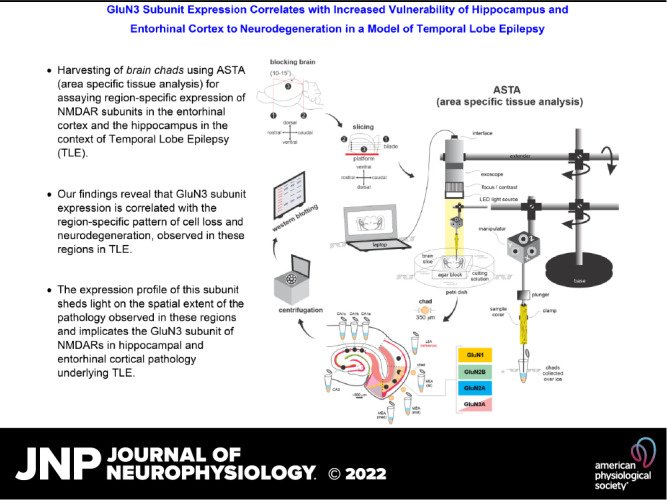
Temporal lobe epilepsy (TLE) is the most common type of epilepsy in adults that is often refractory to antiepileptic medication therapy. Neither the pathology nor the etiology of TLE is fully characterized, although recent studies have established that the two are causally related. TLE pathology entails a stereotypic pattern of neuron loss in hippocampal and parahippocampal regions, predominantly in CA1 subfield of the hippocampus and layer 3 of the medial entorhinal area (MEA), deemed hallmark pathological features of the disease. Through this work, we address the contribution of glutamatergic -methyl-d-aspartate receptors (NMDARs) to the pathology (vulnerability and pattern of neuronal loss), and by extension to the pathophysiology (Ca-induced excitotoxicity), by assaying the spatial expression of their subunit proteins (GluN1, GluN2A, GluN2B, and GluN3A) in these regions using area-specific tissue analysis (ASTA), a novel methodology for harvesting brain chads from hard-to-reach regions within brain slices for Western blotting. Our data suggest gradient expression of the GluN3A subunit along the mid-lateral extent of layer 3 MEA and along the CA1-subicular axis in the hippocampus, unlike GluN1 or GluN2 subunits that are uniformly distributed. Incorporation of GluN3A in the subunit composition of conventional diheteromeric (d-) NMDARs yield triheteromeric (t-) NMDARs which by virtue of their increased selectivity for Ca render neurons vulnerable to excitotoxic damage. Thus, the expression profile of this subunit sheds light on the spatial extent of the pathology observed in these regions and implicates the GluN3 subunit of NMDARs in hippocampal and entorhinal cortical pathology underlying TLE. The role of the GluN3 subunit in NMDAR-mediated pathophysiology underlying TLE is not known. Here, we demonstrate using ASTA (area-specific tissue analysis) that its expression in specific regions of the entorhinal cortex and the hippocampus is correlated with significant cell loss and neurodegeneration, hallmark features of the disease.
颞叶癫痫(TLE)是成人中最常见的癫痫类型,通常对抗癫痫药物治疗有抗性。尽管最近的研究已经确定两者存在因果关系,但 TLE 的病理学和病因仍未完全阐明。TLE 病理学涉及海马和海马旁区域神经元丧失的刻板模式,主要在海马 CA1 亚区和内侧内嗅区(MEA)的第 3 层,被认为是该疾病的标志性病理特征。通过这项工作,我们通过测定这些区域中其亚基蛋白(GluN1、GluN2A、GluN2B 和 GluN3A)的空间表达,来解决谷氨酸 -N- 甲基 -D- 天冬氨酸受体(NMDARs)对病理学(易损性和神经元丧失模式)的贡献,以及通过延伸对生理学(Ca 诱导的兴奋性毒性)的贡献,使用区域特异性组织分析(ASTA),这是一种从脑片中难以到达的区域收获脑块进行 Western blot 的新方法。我们的数据表明,GluN3A 亚基沿 MEA 第 3 层的中外侧延伸以及海马 CA1- 下托轴呈梯度表达,而 GluN1 或 GluN2 亚基则均匀分布。GluN3A 亚基的掺入增加了对 Ca 的选择性,使神经元易受兴奋性毒性损伤,从而形成传统的二聚体(d-)NMDAR 中的三聚体(t-)NMDAR。因此,该亚基的表达谱阐明了这些区域中观察到的病理学的空间范围,并暗示 NMDAR 的 GluN3 亚基参与了 TLE 下的海马和内嗅皮质病理学。GluN3 亚基在 TLE 下 NMDAR 介导的生理学中的作用尚不清楚。在这里,我们使用 ASTA(区域特异性组织分析)证明,其在内嗅皮质和海马特定区域的表达与显著的细胞丢失和神经退行性变相关,这是该疾病的标志性特征。