• 文献检索
  • 文档翻译
  • 深度研究
  • 学术资讯
  • Suppr Zotero 插件Zotero 插件
  • 邀请有礼
  • 套餐&价格
  • 历史记录
应用&插件
Suppr Zotero 插件Zotero 插件浏览器插件Mac 客户端Windows 客户端微信小程序
定价
高级版会员购买积分包购买API积分包
服务
文献检索文档翻译深度研究API 文档MCP 服务
关于我们
关于 Suppr公司介绍联系我们用户协议隐私条款
关注我们

Suppr 超能文献

核心技术专利:CN118964589B侵权必究
粤ICP备2023148730 号-1Suppr @ 2026

文献检索

告别复杂PubMed语法,用中文像聊天一样搜索,搜遍4000万医学文献。AI智能推荐,让科研检索更轻松。

立即免费搜索

文件翻译

保留排版,准确专业,支持PDF/Word/PPT等文件格式,支持 12+语言互译。

免费翻译文档

深度研究

AI帮你快速写综述,25分钟生成高质量综述,智能提取关键信息,辅助科研写作。

立即免费体验

有丝分裂前期微管解体是一个信号提示,它驱动细胞在有丝分裂进入时变圆。

Interphase microtubule disassembly is a signaling cue that drives cell rounding at mitotic entry.

机构信息

Institute for Research in Immunology and Cancer, Université de Montréal, Montréal, Quebec, Canada.

Cellular Mechanisms of Morphogenesis during Mitosis and Cell Motility lab, Université de Montréal, Montréal, Quebec, Canada.

出版信息

J Cell Biol. 2022 Jun 6;221(6). doi: 10.1083/jcb.202109065. Epub 2022 Apr 28.

DOI:10.1083/jcb.202109065
PMID:35482006
原文链接:https://pmc.ncbi.nlm.nih.gov/articles/PMC9059391/
Abstract

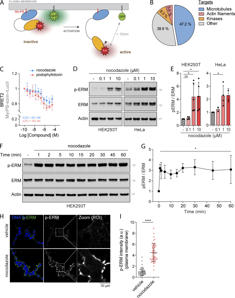
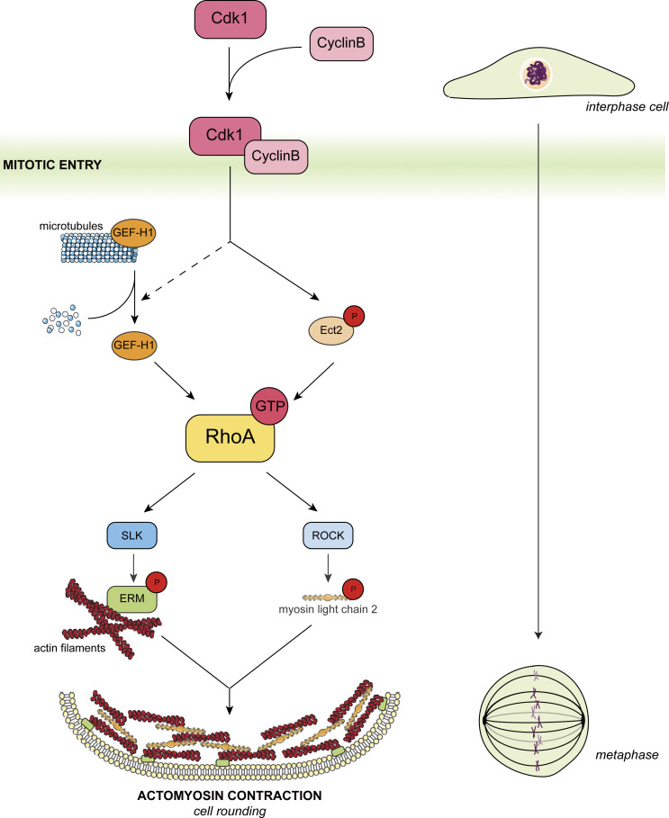
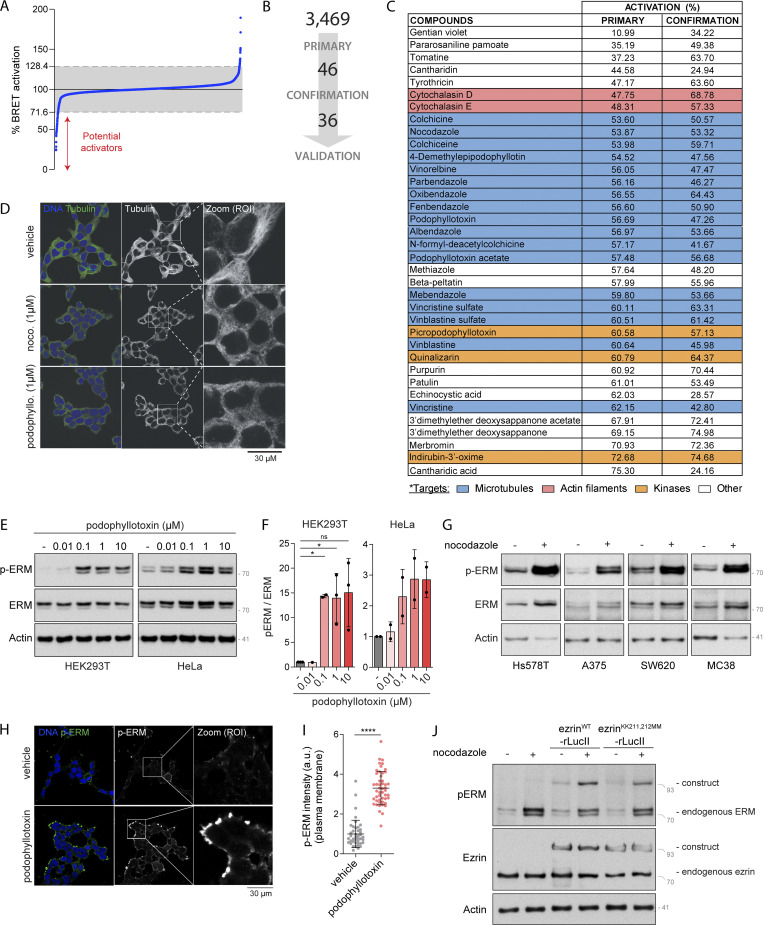
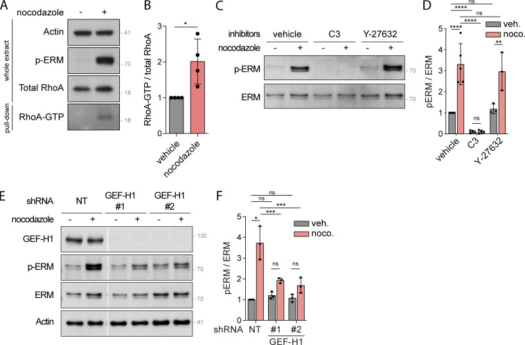
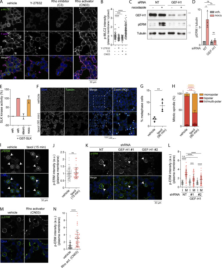
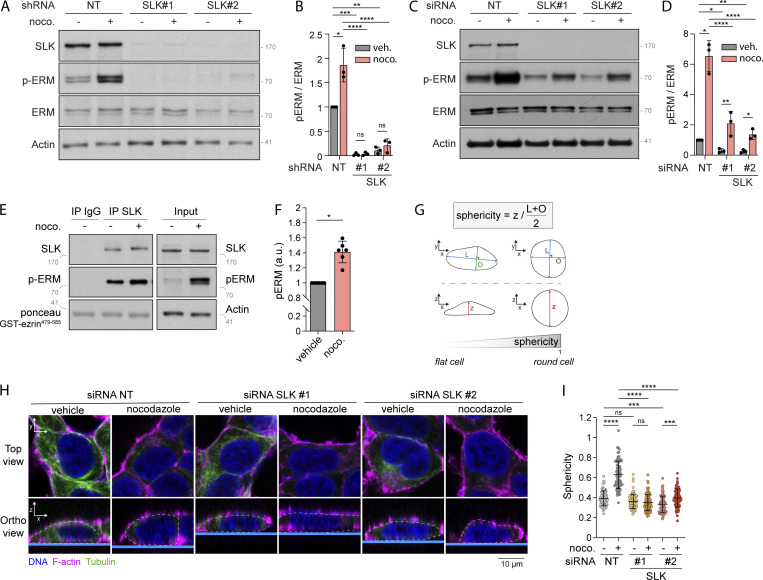
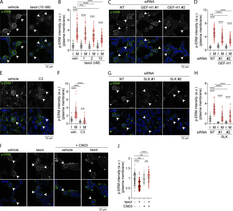
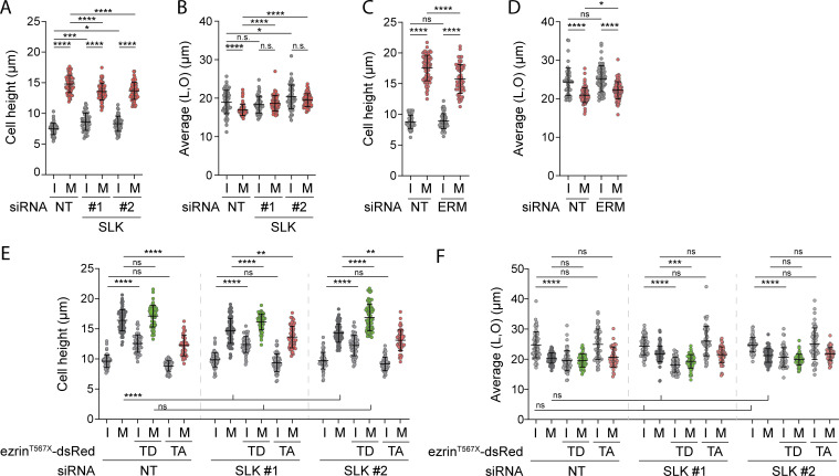
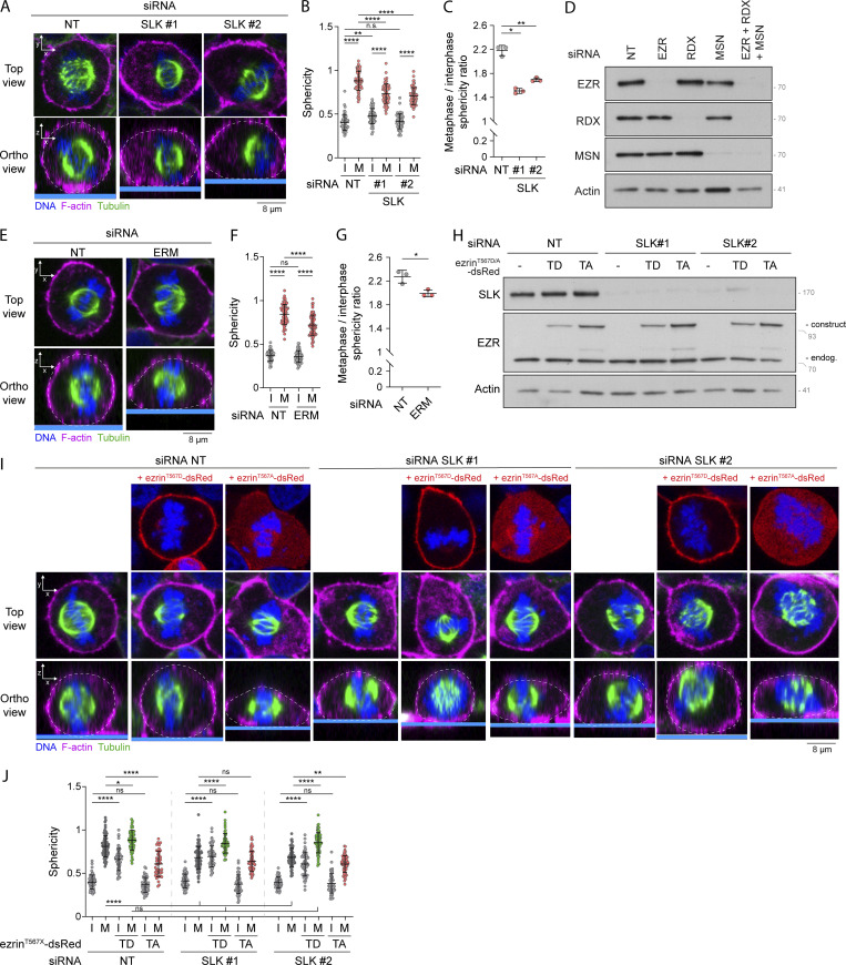
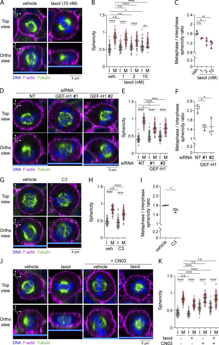
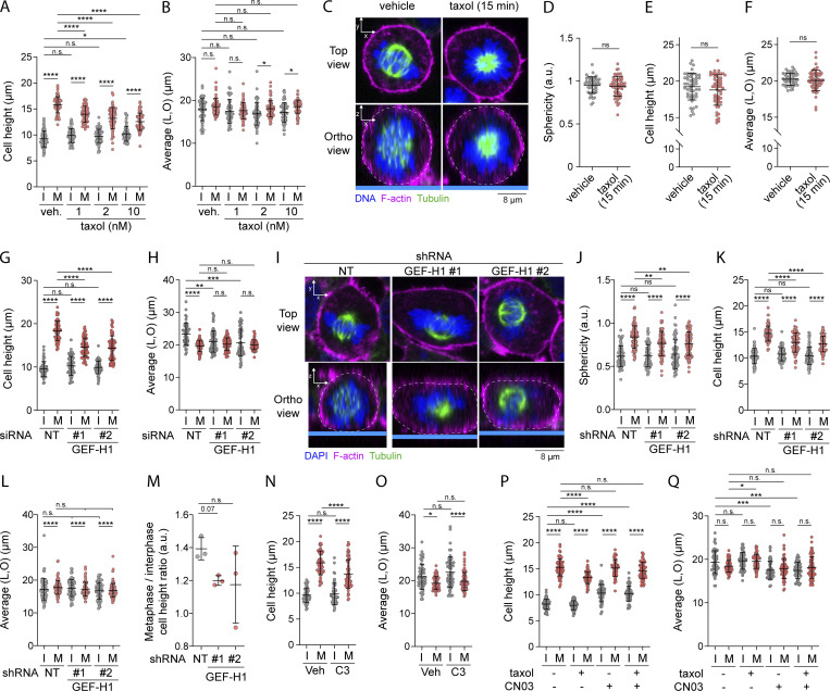
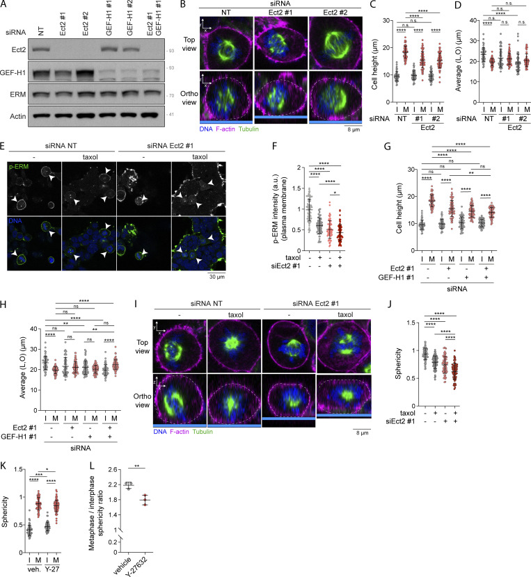
At mitotic entry, reorganization of the actomyosin cortex prompts cells to round-up. Proteins of the ezrin, radixin, and moesin family (ERM) play essential roles in this process by linking actomyosin forces to the plasma membrane. Yet, the cell-cycle signal that activates ERMs at mitotic entry is unknown. By screening a compound library using newly developed biosensors, we discovered that drugs that disassemble microtubules promote ERM activation. We further demonstrated that disassembly of interphase microtubules at mitotic entry directs ERM activation and metaphase cell rounding through GEF-H1, a Rho-GEF inhibited by microtubule binding, RhoA, and its kinase effector SLK. We finally demonstrated that GEF-H1 and Ect2, another Rho-GEF previously identified to control actomyosin forces, act together to drive activation of ERMs and cell rounding in metaphase. In summary, we report microtubule disassembly as a cell-cycle signal that controls a signaling network ensuring that actomyosin forces are efficiently integrated at the plasma membrane to promote cell rounding at mitotic entry.

摘要

在有丝分裂进入时,肌动球蛋白皮层的重新组织促使细胞变圆。埃兹蛋白、radixin 和 moesin 家族(ERM)的蛋白质通过将肌动球蛋白力与质膜连接,在这个过程中发挥着重要作用。然而,激活有丝分裂进入时 ERM 的细胞周期信号尚不清楚。通过使用新开发的生物传感器筛选化合物文库,我们发现解聚微管的药物可促进 ERM 的激活。我们进一步证明,有丝分裂进入时,有丝分裂前期微管的解聚通过 GEF-H1 指导 ERM 的激活和中期细胞变圆,GEF-H1 是一种被微管结合、RhoA 和其激酶效应物 SLK 抑制的 Rho-GEF。我们最终证明,GEF-H1 和 Ect2(另一种先前被确定为控制肌动球蛋白力的 Rho-GEF)共同作用,以驱动 ERM 的激活和中期细胞变圆。总之,我们报告了微管解聚作为细胞周期信号,控制信号网络,确保肌动球蛋白力在质膜上有效整合,以促进有丝分裂进入时的细胞变圆。

https://cdn.ncbi.nlm.nih.gov/pmc/blobs/f0ee/9059391/8cb26750aa70/JCB_202109065_Fig8.jpg
https://cdn.ncbi.nlm.nih.gov/pmc/blobs/f0ee/9059391/2fe13df0e9f7/JCB_202109065_Fig1.jpg
https://cdn.ncbi.nlm.nih.gov/pmc/blobs/f0ee/9059391/e8316398ac7d/JCB_202109065_FigS1.jpg
https://cdn.ncbi.nlm.nih.gov/pmc/blobs/f0ee/9059391/75c11931cb48/JCB_202109065_Fig2.jpg
https://cdn.ncbi.nlm.nih.gov/pmc/blobs/f0ee/9059391/ed3d2a6057d6/JCB_202109065_FigS2.jpg
https://cdn.ncbi.nlm.nih.gov/pmc/blobs/f0ee/9059391/f00c26cd85f1/JCB_202109065_Fig3.jpg
https://cdn.ncbi.nlm.nih.gov/pmc/blobs/f0ee/9059391/9f25737970ab/JCB_202109065_Fig4.jpg
https://cdn.ncbi.nlm.nih.gov/pmc/blobs/f0ee/9059391/6928f4ff82a5/JCB_202109065_FigS3.jpg
https://cdn.ncbi.nlm.nih.gov/pmc/blobs/f0ee/9059391/7ed87d9ec742/JCB_202109065_Fig5.jpg
https://cdn.ncbi.nlm.nih.gov/pmc/blobs/f0ee/9059391/8425c1d0c115/JCB_202109065_Fig6.jpg
https://cdn.ncbi.nlm.nih.gov/pmc/blobs/f0ee/9059391/e27325b7462f/JCB_202109065_FigS4.jpg
https://cdn.ncbi.nlm.nih.gov/pmc/blobs/f0ee/9059391/91e262830215/JCB_202109065_Fig7.jpg
https://cdn.ncbi.nlm.nih.gov/pmc/blobs/f0ee/9059391/22ddccd96bec/JCB_202109065_FigS5.jpg
https://cdn.ncbi.nlm.nih.gov/pmc/blobs/f0ee/9059391/8cb26750aa70/JCB_202109065_Fig8.jpg

相似文献

1
Interphase microtubule disassembly is a signaling cue that drives cell rounding at mitotic entry.有丝分裂前期微管解体是一个信号提示,它驱动细胞在有丝分裂进入时变圆。
J Cell Biol. 2022 Jun 6;221(6). doi: 10.1083/jcb.202109065. Epub 2022 Apr 28.
2
BNIP-2 retards breast cancer cell migration by coupling microtubule-mediated GEF-H1 and RhoA activation.BNIP-2 通过耦联微管介导的 GEF-H1 和 RhoA 激活来抑制乳腺癌细胞迁移。
Sci Adv. 2020 Jul 31;6(31):eaaz1534. doi: 10.1126/sciadv.aaz1534. eCollection 2020 Jul.
3
Changes in Ect2 localization couple actomyosin-dependent cell shape changes to mitotic progression.Ect2 定位的变化将依赖肌动球蛋白的细胞形状变化与有丝分裂进程偶联。
Dev Cell. 2012 Aug 14;23(2):371-83. doi: 10.1016/j.devcel.2012.06.003.
4
Reorganization of paclitaxel-stabilized microtubule arrays at mitotic entry: roles of depolymerizing kinesins and severing proteins.有丝分裂起始时紫杉醇稳定的微管阵列的重排:解聚驱动蛋白和切割蛋白的作用。
Cancer Biol Ther. 2019;20(10):1337-1347. doi: 10.1080/15384047.2019.1638678. Epub 2019 Jul 25.
5
Spatiotemporal dynamics of GEF-H1 activation controlled by microtubule- and Src-mediated pathways.微管和Src 介导的途径控制 GEF-H1 激活的时空动力学。
J Cell Biol. 2019 Sep 2;218(9):3077-3097. doi: 10.1083/jcb.201812073. Epub 2019 Aug 16.
6
The actin-binding ERM protein Moesin binds to and stabilizes microtubules at the cell cortex.细胞皮层处肌动蛋白结合型 ERM 蛋白 Moesin 与微管结合并使其稳定。
J Cell Biol. 2013 Jul 22;202(2):251-60. doi: 10.1083/jcb.201304052. Epub 2013 Jul 15.
7
Role of guanine nucleotide exchange factor-H1 in complement-mediated RhoA activation in glomerular epithelial cells.鸟嘌呤核苷酸交换因子-H1 在补体介导的肾小球上皮细胞 RhoA 激活中的作用。
J Biol Chem. 2014 Feb 14;289(7):4206-18. doi: 10.1074/jbc.M113.506816. Epub 2013 Dec 19.
8
Regulation and functions of the RhoA regulatory guanine nucleotide exchange factor GEF-H1.RhoA 调节性鸟嘌呤核苷酸交换因子 GEF-H1 的调节和功能。
Small GTPases. 2021 Sep-Nov;12(5-6):358-371. doi: 10.1080/21541248.2020.1840889. Epub 2020 Oct 30.
9
Guanine nucleotide exchange factor-H1 regulates cell migration via localized activation of RhoA at the leading edge.鸟嘌呤核苷酸交换因子-H1通过在前缘局部激活RhoA来调节细胞迁移。
Mol Biol Cell. 2009 Sep;20(18):4070-82. doi: 10.1091/mbc.e09-01-0041. Epub 2009 Jul 22.
10
Hydrostatic pressure and the actomyosin cortex drive mitotic cell rounding.静水压和肌动球蛋白皮层驱动有丝分裂细胞的圆形化。
Nature. 2011 Jan 13;469(7329):226-30. doi: 10.1038/nature09642. Epub 2011 Jan 2.

引用本文的文献

1
A Benzodiazepine-Derived Molecule That Interferes with the Bio-Mechanical Properties of Glioblastoma-Astrocytoma Cells Altering Their Proliferation and Migration.一种干扰胶质母细胞瘤-星形细胞瘤细胞生物力学特性、改变其增殖和迁移的苯二氮卓类衍生分子。
Int J Mol Sci. 2025 Mar 19;26(6):2767. doi: 10.3390/ijms26062767.
2
Slik sculpts the plasma membrane into cytonemes to control cell-cell communication.Slik将质膜塑造为丝状伪足以控制细胞间通讯。
EMBO J. 2025 Apr;44(8):2186-2210. doi: 10.1038/s44318-025-00401-8. Epub 2025 Mar 6.
3
Dynamic Coupling of MAPK Signaling to the Guanine Nucleotide Exchange Factor GEF-H1.

本文引用的文献

1
Development of conformational BRET biosensors that monitor ezrin, radixin and moesin activation in real time.开发构象型 BRET 生物传感器,实时监测埃兹蛋白、根蛋白和膜突蛋白的激活。
J Cell Sci. 2021 Apr 1;134(7). doi: 10.1242/jcs.255307. Epub 2021 Apr 13.
2
Regulation and functions of the RhoA regulatory guanine nucleotide exchange factor GEF-H1.RhoA 调节性鸟嘌呤核苷酸交换因子 GEF-H1 的调节和功能。
Small GTPases. 2021 Sep-Nov;12(5-6):358-371. doi: 10.1080/21541248.2020.1840889. Epub 2020 Oct 30.
3
STRIPAK regulates Slik localization to control mitotic morphogenesis and epithelial integrity.
丝裂原活化蛋白激酶(MAPK)信号与鸟嘌呤核苷酸交换因子GEF-H1的动态偶联
Onco Targets Ther. 2025 Jan 25;18:147-159. doi: 10.2147/OTT.S496228. eCollection 2025.
4
Regulation of epithelial cell jamming transition by cytoskeleton and cell-cell interactions.细胞骨架和细胞间相互作用对上皮细胞阻塞转变的调控。
Biophys Rev (Melville). 2024 Oct 14;5(4):041301. doi: 10.1063/5.0220088. eCollection 2024 Dec.
5
YAP Overcomes Mechanical Barriers to Induce Mitotic Rounding and Adult Cardiomyocyte Division.YAP克服机械屏障以诱导有丝分裂变圆和成年心肌细胞分裂。
Circulation. 2025 Jan 7;151(1):76-93. doi: 10.1161/CIRCULATIONAHA.123.066004. Epub 2024 Oct 11.
6
Excitable Rho dynamics control cell shape and motility by sequentially activating ERM proteins and actomyosin contractility.激动的 Rho 动力学通过顺序激活 ERM 蛋白和肌动球蛋白收缩来控制细胞形状和运动性。
Sci Adv. 2024 Sep 6;10(36):eadn6858. doi: 10.1126/sciadv.adn6858.
7
Ezrin, radixin, and moesin are dispensable for macrophage migration and cellular cortex mechanics.埃兹蛋白、根蛋白和膜突蛋白对于巨噬细胞迁移和细胞皮质力学是可有可无的。
EMBO J. 2024 Nov;43(21):4822-4845. doi: 10.1038/s44318-024-00173-7. Epub 2024 Jul 18.
8
Osmotic Pressure and Its Biological Implications.渗透压力及其生物学意义。
Int J Mol Sci. 2024 Mar 14;25(6):3310. doi: 10.3390/ijms25063310.
9
Rac1 promotes kidney collecting duct repair by mechanically coupling cell morphology to mitotic entry.Rac1 通过机械偶联细胞形态与有丝分裂进入来促进肾集合管修复。
Sci Adv. 2024 Feb 9;10(6):eadi7840. doi: 10.1126/sciadv.adi7840. Epub 2024 Feb 7.
10
Bone resorption by osteoclasts involves fine tuning of RHOA activity by its microtubule-associated exchange factor GEF-H1.破骨细胞介导的骨吸收涉及通过其微管相关交换因子GEF-H1对RHOA活性进行精细调节。
Front Physiol. 2024 Jan 19;15:1342024. doi: 10.3389/fphys.2024.1342024. eCollection 2024.
STRIPAK 调控 Slik 的定位以控制有丝分裂形态发生和上皮完整性。
J Cell Biol. 2020 Nov 2;219(11). doi: 10.1083/jcb.201911035.
4
The Mechanics of Mitotic Cell Rounding.有丝分裂细胞变圆的机制。
Front Cell Dev Biol. 2020 Aug 6;8:687. doi: 10.3389/fcell.2020.00687. eCollection 2020.
5
Oncogenic Signaling Alters Cell Shape and Mechanics to Facilitate Cell Division under Confinement.致癌信号改变细胞形状和力学特性,以促进受限环境下的细胞分裂。
Dev Cell. 2020 Mar 9;52(5):563-573.e3. doi: 10.1016/j.devcel.2020.01.004. Epub 2020 Feb 6.
6
Mapping the proximity interaction network of the Rho-family GTPases reveals signalling pathways and regulatory mechanisms.绘制 Rho 家族 GTP 酶的邻近相互作用网络图谱揭示了信号通路和调控机制。
Nat Cell Biol. 2020 Jan;22(1):120-134. doi: 10.1038/s41556-019-0438-7. Epub 2019 Dec 23.
7
Context is everything: aneuploidy in cancer.背景至关重要:癌症中的非整倍体。
Nat Rev Genet. 2020 Jan;21(1):44-62. doi: 10.1038/s41576-019-0171-x. Epub 2019 Sep 23.
8
Spatiotemporal dynamics of GEF-H1 activation controlled by microtubule- and Src-mediated pathways.微管和Src 介导的途径控制 GEF-H1 激活的时空动力学。
J Cell Biol. 2019 Sep 2;218(9):3077-3097. doi: 10.1083/jcb.201812073. Epub 2019 Aug 16.
9
Reorganization of paclitaxel-stabilized microtubule arrays at mitotic entry: roles of depolymerizing kinesins and severing proteins.有丝分裂起始时紫杉醇稳定的微管阵列的重排:解聚驱动蛋白和切割蛋白的作用。
Cancer Biol Ther. 2019;20(10):1337-1347. doi: 10.1080/15384047.2019.1638678. Epub 2019 Jul 25.
10
A mechano-signalling network linking microtubules, myosin IIA filaments and integrin-based adhesions.一个机械信号网络,将微管、肌球蛋白 IIA 丝和整合素基黏附连接起来。
Nat Mater. 2019 Jun;18(6):638-649. doi: 10.1038/s41563-019-0371-y. Epub 2019 May 21.