Neurological Surgery Research Laboratory, Department of Neurosurgery, Montefiore Medical Center and Albert Einstein College of Medicine, Bronx, NY, USA.
Department of Pathology, Albert Einstein College of Medicine, Bronx, NY, USA.
Sci Rep. 2022 Apr 28;12(1):6934. doi: 10.1038/s41598-022-10793-w.
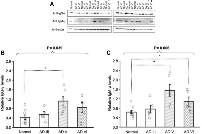
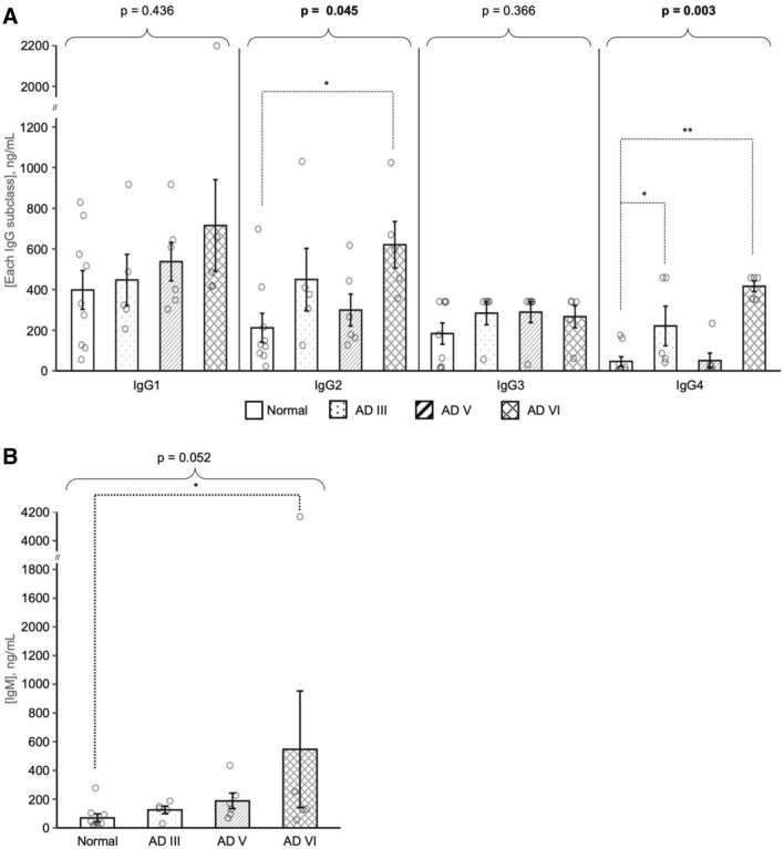
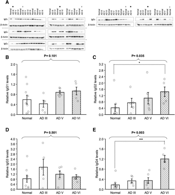
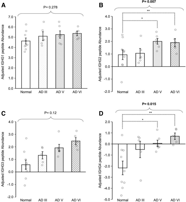
The immune system has been described to play a role in the development of Alzheimer's disease (AD), but the distribution of immunoglobulins and their subclasses in brain tissue has not been explored. In this study, examination of pathologically diagnosed frontal cortex gray matter revealed significantly higher levels of IgM and IgG in late-stage AD (Braak and Braak stages V and VI) compared to age-matched controls. While levels of IgG2 and IgG4 constant region fragments were higher in late-stage AD, concentration of native-state IgG4 with free Fc regions was increased in AD III and VI. RNA analysis did not support parenchymal B-cell production of IgG4 in AD III and V, indicating possible peripheral or meningeal B-cell involvement. Changes in the profile of IgM, IgG and IgG subclasses in AD frontal cortex may provide insight into understanding disease pathogenesis and progression.
免疫系统被描述为在阿尔茨海默病 (AD) 的发展中起作用,但免疫球蛋白及其亚型在脑组织中的分布尚未得到探索。在这项研究中,对病理诊断的额皮质灰质的检查显示,与年龄匹配的对照组相比,晚期 AD(Braak 和 Braak 阶段 V 和 VI)中 IgM 和 IgG 的水平显著升高。虽然晚期 AD 中 IgG2 和 IgG4 恒定区片段的水平较高,但 AD III 和 VI 中具有游离 Fc 区的天然状态 IgG4 的浓度增加。RNA 分析不支持 AD III 和 V 中实质 B 细胞产生 IgG4,表明可能涉及外周或脑膜 B 细胞。AD 额皮质中 IgM、IgG 和 IgG 亚型谱的变化可能有助于了解疾病的发病机制和进展。