Department of Otorhinolaryngology and Head-and-Neck Surgery, Akita University Graduate School of Medicine, Akita, 010-8543 Japan.
Department of Molecular and Tumour Pathology, Akita University Graduate School of Medicine, Akita, 010-8543 Japan.
Asian Pac J Cancer Prev. 2022 Apr 1;23(4):1271-1278. doi: 10.31557/APJCP.2022.23.4.1271.
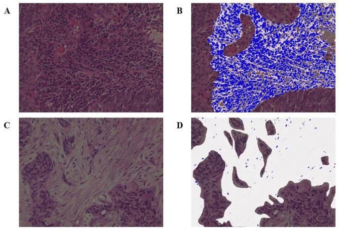
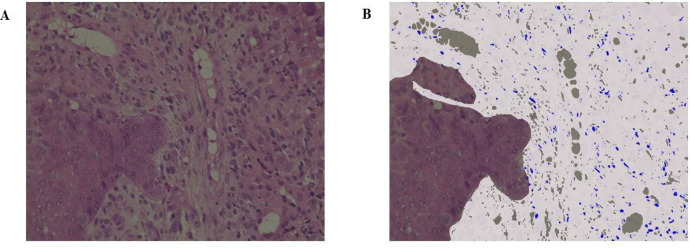
Tumor-infiltrating lymphocytes (TILs) are assessed by the ratio of the area of lymphocytes infiltrating the stroma. TILs are important in breast cancer and malignant melanoma and are being established as a marker of prognosis and sensitivity to chemotherapy. This has resulted in various therapies being developed in fields such as breast cancer. However, the evaluation of TILs in head and neck squamous cell carcinoma (HNSCC) is not progressing, and the prognosis is still poor. Thus, investigating whether or not the evaluation of TILs is also effective in HNSCC and prognoses can be predicted with just biopsy samples alone is required.
This study included 153 patients who were diagnosed with HNSCC between January 2010 and December 2019, underwent treatment, and could be followed up thereafter at our institution.
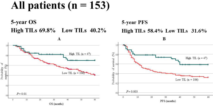
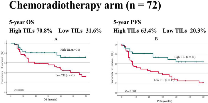
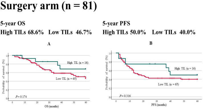
TILs, overall survival (OS), and progression-free survival (PFS) were evaluated in all patients, the chemoradiotherapy arm, and the surgery arm. The cut-off value for TILs was 50%. In all patients, OS was 69.8% and 40.2% (P = 0.01) and PFS was 58.4% and 31.6% (P = 0.003) in the high and low TIL groups, respectively. Multivariate analyses revealed that TILs independently predicted prognosis. In the chemoradiotherapy arm, OS was 70.8% and 31.6% (P = 0.012) and PFS was 63.4% and 20.3% (P = 0.001) in the high and low TIL groups, respectively. No significant differences were noted in the surgery arm.
In HNSCC, TILs can be used as a prognosis predictor and chemoradiotherapy biomarker. Assessments can be performed just with hematoxylin-eosin staining and is very simple. This will greatly contribute to report personalized therapy progress. Further evaluations and, thus, prospective clinical multicenter trials are needed to use TILs in clinical practice for HNSCC.
肿瘤浸润淋巴细胞(TILs)通过浸润间质的淋巴细胞面积比来评估。TILs 在乳腺癌和恶性黑色素瘤中很重要,并且被确立为预后和对化疗敏感性的标志物。这导致在乳腺癌等领域开发了各种疗法。然而,头颈部鳞状细胞癌(HNSCC)中 TILs 的评估没有进展,预后仍然很差。因此,需要研究仅通过活检样本评估 TILs 在 HNSCC 中是否也有效,以及是否可以预测预后。
本研究纳入了 2010 年 1 月至 2019 年 12 月期间在我院诊断为 HNSCC、接受治疗并可随后进行随访的 153 例患者。
所有患者、放化疗组和手术组均评估了 TILs、总生存期(OS)和无进展生存期(PFS)。TILs 的截断值为 50%。在所有患者中,TILs 高表达组的 OS 为 69.8%和 40.2%(P = 0.01),PFS 为 58.4%和 31.6%(P = 0.003),低表达组分别为 58.4%和 31.6%。多变量分析表明 TILs 独立预测预后。在放化疗组中,TILs 高表达组的 OS 为 70.8%和 31.6%(P = 0.012),PFS 为 63.4%和 20.3%(P = 0.001),低表达组分别为 63.4%和 20.3%。手术组无显著差异。
在 HNSCC 中,TILs 可用作预后预测和放化疗的生物标志物。仅通过苏木精-伊红染色即可进行评估,非常简单。这将极大地促进报告个性化治疗进展。需要进一步评估,因此需要前瞻性的临床多中心试验,以便在 HNSCC 的临床实践中使用 TILs。