Eye Institute of Xiamen University, Fujian Provincial Key Laboratory of Ophthalmology and Visual Science, School of Medicine, Xiamen University, Xiamen, Fujian 361104, China; Department of Ophthalmology, Massachusetts Eye and Ear, Harvard Medical School, Boston, Massachusetts 02114, USA.
Eye Institute of Xiamen University, Fujian Provincial Key Laboratory of Ophthalmology and Visual Science, School of Medicine, Xiamen University, Xiamen, Fujian 361104, China; Department of Ophthalmology, Zhongshan Hospital of Xiamen University, School of Medicine, Xiamen University, Xiamen, Fujian 361004, China.
Stem Cell Reports. 2022 May 10;17(5):1105-1119. doi: 10.1016/j.stemcr.2022.03.017. Epub 2022 Apr 28.
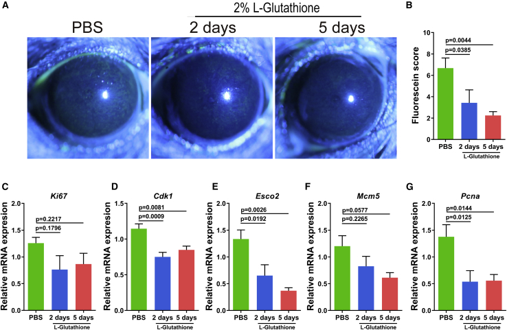
Sleep deficiency, a common public health problem, causes ocular discomfort and affects ocular surface health. However, the underlying mechanism remains unclear. Herein, we identified that short-term sleep deprivation (SD) resulted in hyperproliferation of corneal epithelial progenitor cells (CEPCs) in mice. The expression levels of p63 and Keratin 14, the biomarkers of CEPCs, were upregulated in the corneal epithelium after short-term SD. In addition, SD led to elevated levels of reactive oxygen species (ROS), and subsequent decrease in antioxidant capacity, in the tear film. Exogenous hydrogen peroxide (HO) could directly stimulate the proliferation of CEPCs in vivo and in vitro. Topical treatment of antioxidant L-glutathione preserved the over-proliferation of CEPCs and attenuated corneal epithelial defects in SD mice. Moreover, the activation of the phosphoinositide 3-kinase (PI3K)/AKT signaling pathway is essential to ROS-stimulated cell proliferation in CEPCs. However, long-term SD ultimately led to early manifestation of limbal stem cell deficiency.
睡眠不足是一种常见的公共卫生问题,它会引起眼部不适,并影响眼表面健康。然而,其潜在的机制尚不清楚。在此,我们发现短期睡眠剥夺会导致小鼠角膜上皮祖细胞(CEPC)过度增殖。短期 SD 后,角膜上皮中 p63 和角蛋白 14(CEPC 的生物标志物)的表达水平上调。此外,SD 导致泪膜中活性氧(ROS)水平升高,随后抗氧化能力下降。外源性过氧化氢(HO)可直接刺激 CEPC 的体内和体外增殖。局部抗氧化剂 L-谷胱甘肽治疗可维持 CEPC 的过度增殖,并减轻 SD 小鼠的角膜上皮缺损。此外,PI3K/AKT 信号通路的激活对于 ROS 刺激的 CEPC 增殖至关重要。然而,长期 SD 最终导致角膜缘干细胞缺乏的早期表现。