das Virgens Aquino Maria Jane, Dos Santos Leite Paula Michele, Lima Rodrigues Ingrid Kyelli, DeSantana Josimari Melo
Physiotherapy Department, Federal University of Sergipe, São Cristóvão, Brazil.
Physical Therapy Department, Graduate Program in Health Science, Graduate Program in Physiological Science, Federal University of Sergipe, São Cristóvão, Brazil.
Front Oncol. 2022 Apr 14;12:740787. doi: 10.3389/fonc.2022.740787. eCollection 2022.
Breast cancer is the most common in the female population. Physical training is safe and indicated after surgical treatment for breast cancer. During exercise, body temperature changes due to tissue metabolic activity; in this sense, infrared thermography is used to map the thermal patterns of the body surface.
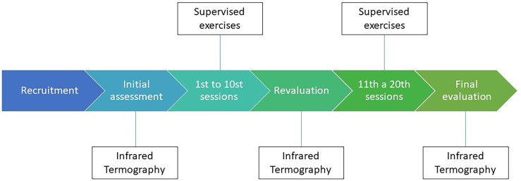
This study aimed to evaluate the feasibility of using thermography during a physical rehabilitation program in mastectomized patients by analyzing the change in body temperature caused by physical exercise in the breast region.
This is a simple and covert clinical trial, in which the sample was constituted for convenience. The women were submitted to a supervised physical exercise protocol, three times a week, for 20 sessions. They were evaluated in the first, tenth, and twentieth sessions in relation to changes in body temperature in the breast region (infrared thermography).
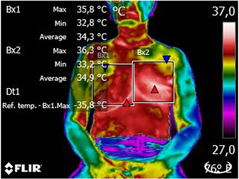
Twenty patients who underwent mastectomy surgery were recruited. No patient had drain infection, scar dehiscence, or lymphedema, and only one patient had seroma removed. The mean age was 50.45 ± 2.00 years, and the body mass index (BMI) was 28.95 ± 1.11 kg/m. In the body thermography of the patients' breast region, no significant difference was observed when comparing the thermograms of the plastron region of the patients in the first, tenth, and twentieth sessions (p = 0.201). However, when comparing the plastron region with the control breast, a reduction in temperature was observed in the operated region in the first (p = 0.012) and tenth sessions (p = 0.004).
Through this study, we can conclude that the use of infrared thermography is viable for the analysis of the body temperature of mastectomized patients during a supervised physical exercise protocol and, therefore, suggest that this instrument is increasingly used in the cancer public.
乳腺癌是女性群体中最常见的癌症。体育锻炼对乳腺癌手术后的患者来说是安全且适宜的。在运动过程中,体温会因组织代谢活动而发生变化;从这个意义上讲,红外热成像技术被用于绘制体表的热模式。
本研究旨在通过分析体育锻炼引起的乳房区域体温变化,评估热成像技术在乳房切除术后患者的物理康复计划中的可行性。
这是一项简单且隐蔽的临床试验,样本选取基于便利性。女性患者接受有监督的体育锻炼方案,每周三次,共20节课程。在第1、10和20节课程中,对她们乳房区域的体温变化(红外热成像)进行评估。
招募了20名接受乳房切除术的患者。没有患者出现引流感染、切口裂开或淋巴水肿,只有一名患者进行了血清肿清除。平均年龄为50.45±2.00岁,体重指数(BMI)为28.95±1.11kg/m。在患者乳房区域的身体热成像中,比较第1、10和20节课程中患者胸骨区域的热图时,未观察到显著差异(p = 0.201)。然而,将胸骨区域与对照乳房进行比较时,在第1节(p = 0.012)和第10节(p = 0.004)课程中,手术区域的温度出现了降低。
通过本研究,我们可以得出结论,在有监督的体育锻炼方案中,使用红外热成像技术分析乳房切除术后患者的体温是可行的,因此建议该仪器在癌症患者群体中得到更广泛的应用。