Brunner Clément, Macé Emilie, Montaldo Gabriel, Urban Alan
Neuro-Electronics Research Flanders, Leuven, Belgium.
VIB, Leuven, Belgium.
Front Neurosci. 2022 Apr 12;16:831650. doi: 10.3389/fnins.2022.831650. eCollection 2022.
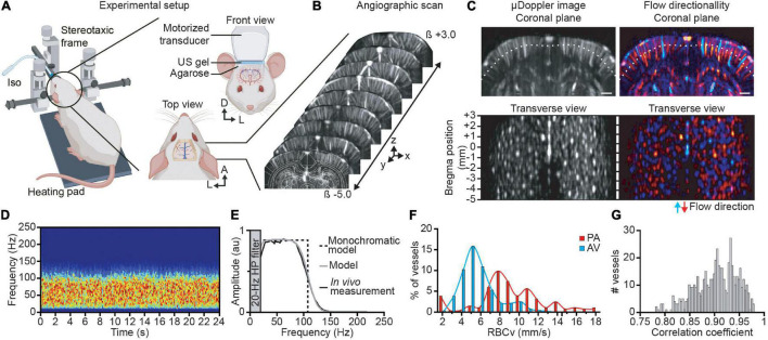
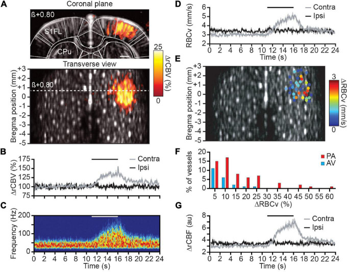
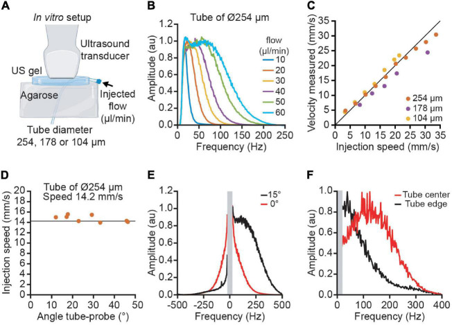
Red blood cell velocity (RBCv), cerebral blood flow (CBF), and volume (CBV) are three key parameters when describing brain hemodynamics. Functional ultrasound imaging is a Doppler-based method allowing for real-time measurement of relative CBV at high spatiotemporal resolution (100 × 110 × 300 μm, up to 10 Hz) and large scale. Nevertheless, the measure of RBCv and CBF in small cortical vessels with functional ultrasound imaging remains challenging because of their orientation and size, which impairs the ability to perform precise measurements. We designed a directional flow filter to overpass these limitations allowing us to measure RBCv in single vessels using a standard functional ultrasound imaging system without contrast agents (e.g., microbubbles). This method allows to quickly extract the number of vessels in the cortex that was estimated to be approximately 650/cm in adult rats, with a 55-45% ratio for penetrating arterioles versus ascending venules. Then, we analyzed the changes in RBCv in these vessels during forepaw stimulation. We observed that ∼40 vessels located in the primary somatosensory forelimb cortex display a significant increase of the RBCv (median ΔRBCv ∼15%, maximal ΔRBCv ∼60%). As expected, we show that RBCv was higher for penetrating arterioles located in the center than in the periphery of the activated area. The proposed approach extends the capabilities of functional ultrasound imaging, which may contribute to a better understanding of the neurovascular coupling at the brain-wide scale.
红细胞速度(RBCv)、脑血流量(CBF)和脑血容量(CBV)是描述脑血流动力学的三个关键参数。功能超声成像基于多普勒原理,能够以高时空分辨率(100×110×300μm,最高可达10Hz)大规模实时测量相对脑血容量。然而,由于小皮质血管的方向和大小,使用功能超声成像测量其红细胞速度和脑血流量仍然具有挑战性,这影响了精确测量的能力。我们设计了一种定向血流滤波器来克服这些限制,使我们能够使用无造影剂(如微泡)的标准功能超声成像系统测量单个血管中的红细胞速度。该方法能够快速提取皮质中的血管数量,在成年大鼠中估计约为650根/cm,其中穿通小动脉与上行小静脉的比例为55-45%。然后,我们分析了前爪刺激期间这些血管中红细胞速度的变化。我们观察到,位于初级体感前肢皮质的约40根血管的红细胞速度显著增加(红细胞速度中位数变化约15%,最大变化约60%)。正如预期的那样,我们发现位于激活区域中心的穿通小动脉的红细胞速度高于周边区域。所提出的方法扩展了功能超声成像的能力,这可能有助于在全脑范围内更好地理解神经血管耦合。