• 文献检索
  • 文档翻译
  • 深度研究
  • 学术资讯
  • Suppr Zotero 插件Zotero 插件
  • 邀请有礼
  • 套餐&价格
  • 历史记录
应用&插件
Suppr Zotero 插件Zotero 插件浏览器插件Mac 客户端Windows 客户端微信小程序
定价
高级版会员购买积分包购买API积分包
服务
文献检索文档翻译深度研究API 文档MCP 服务
关于我们
关于 Suppr公司介绍联系我们用户协议隐私条款
关注我们

Suppr 超能文献

核心技术专利:CN118964589B侵权必究
粤ICP备2023148730 号-1Suppr @ 2026

文献检索

告别复杂PubMed语法,用中文像聊天一样搜索,搜遍4000万医学文献。AI智能推荐,让科研检索更轻松。

立即免费搜索

文件翻译

保留排版,准确专业,支持PDF/Word/PPT等文件格式,支持 12+语言互译。

免费翻译文档

深度研究

AI帮你快速写综述,25分钟生成高质量综述,智能提取关键信息,辅助科研写作。

立即免费体验

纳米金刚石聚集体的结构演变:小角中子散射和超小角中子散射研究

Structure evolution of nanodiamond aggregates: a SANS and USANS study.

作者信息

Kabir Imrana I, Osborn John C, Lu Weijian, Mata Jitendra P, Rehm Christine, Yeoh Guan H, Ersez Tunay

机构信息

School of Mechanical and Manufacturing Engineering, University of New South Wales, Sydney, NSW 2052, Australia.

Australian Nuclear Science and Technology Organisation, Locked Bag 2001, Kirrawee DC, NSW 2232, Australia.

出版信息

J Appl Crystallogr. 2022 Mar 25;55(Pt 2):353-361. doi: 10.1107/S1600576722002084. eCollection 2022 Apr 1.

DOI:10.1107/S1600576722002084
PMID:35497657
原文链接:https://pmc.ncbi.nlm.nih.gov/articles/PMC8985605/
Abstract

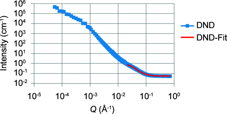
Ultra-small-angle neutron scattering (USANS) and small-angle neutron scattering (SANS) measurements, covering length scales from micrometres to nanometres, were made to investigate the structure of nanodiamonds (NDs) and their suspensions. These nanodiamonds were produced by two different techniques, namely by the detonation method and by the laser ablation of a carbon-hydro-carbon mixture. The (U)SANS results indicated the presence of structures four orders of magnitude larger than the dimensions of a single ND particle, consisting of aggregations of ND particles. This aggregation of the ND particles was studied by employing the contrast variation technique. Two different solvents, namely HO and dimethyl sulfoxide (and their deuterated counterparts), were used to understand the role of hydrogen in the shape and size of the aggregates. The analysis of experimental data from SANS measurements also reveals the ND particles to have an ellipsoidal structure. Using a defined shape model and the SANS contrast variation technique, it was possible to characterize the non-diamond outer shell of the particles and determine the outer layer thickness. This clarification of the structure of the NDs will allow better preparation of suspensions/samples for various applications. Understanding the structure of NDs at multiple length scales also provides crucial knowledge of particle-particle interaction and its effect on the aggregation structures.

摘要

为了研究纳米金刚石(NDs)及其悬浮液的结构,进行了超小角中子散射(USANS)和小角中子散射(SANS)测量,测量范围覆盖从微米到纳米的长度尺度。这些纳米金刚石是通过两种不同技术制备的,即爆轰法和碳 - 碳氢混合物的激光烧蚀法。(U)SANS结果表明存在比单个ND颗粒尺寸大四个数量级的结构,该结构由ND颗粒的聚集体组成。通过采用对比变化技术研究了ND颗粒的这种聚集现象。使用两种不同的溶剂,即水和二甲基亚砜(及其氘代对应物)来了解氢在聚集体形状和尺寸中的作用。对SANS测量实验数据的分析还揭示了ND颗粒具有椭圆形结构。使用定义的形状模型和SANS对比变化技术,可以表征颗粒的非金刚石外壳并确定外层厚度。对NDs结构的这种阐明将有助于更好地制备用于各种应用的悬浮液/样品。了解多个长度尺度上的NDs结构还提供了关于颗粒 - 颗粒相互作用及其对聚集结构影响的关键知识。

https://cdn.ncbi.nlm.nih.gov/pmc/blobs/3aef/8985605/3ef81884f05f/j-55-00353-fig9.jpg
https://cdn.ncbi.nlm.nih.gov/pmc/blobs/3aef/8985605/7ff4755b28d6/j-55-00353-fig1.jpg
https://cdn.ncbi.nlm.nih.gov/pmc/blobs/3aef/8985605/dbafbdcadb94/j-55-00353-fig2.jpg
https://cdn.ncbi.nlm.nih.gov/pmc/blobs/3aef/8985605/6ad0115abbe0/j-55-00353-fig3.jpg
https://cdn.ncbi.nlm.nih.gov/pmc/blobs/3aef/8985605/6be97b12ecb0/j-55-00353-fig4.jpg
https://cdn.ncbi.nlm.nih.gov/pmc/blobs/3aef/8985605/0c8c4ab73431/j-55-00353-fig5.jpg
https://cdn.ncbi.nlm.nih.gov/pmc/blobs/3aef/8985605/38377ddcb08e/j-55-00353-fig6.jpg
https://cdn.ncbi.nlm.nih.gov/pmc/blobs/3aef/8985605/247308818d9a/j-55-00353-fig7.jpg
https://cdn.ncbi.nlm.nih.gov/pmc/blobs/3aef/8985605/aede60d83933/j-55-00353-fig8.jpg
https://cdn.ncbi.nlm.nih.gov/pmc/blobs/3aef/8985605/3ef81884f05f/j-55-00353-fig9.jpg

相似文献

1
Structure evolution of nanodiamond aggregates: a SANS and USANS study.纳米金刚石聚集体的结构演变:小角中子散射和超小角中子散射研究
J Appl Crystallogr. 2022 Mar 25;55(Pt 2):353-361. doi: 10.1107/S1600576722002084. eCollection 2022 Apr 1.
2
Effects of soil particles and convective transport on dispersion and aggregation of nanoplastics via small-angle neutron scattering (SANS) and ultra SANS (USANS).通过小角中子散射(SANS)和超小角中子散射(USANS)研究土壤颗粒和对流输运对纳米塑料分散和聚集的影响。
PLoS One. 2020 Jul 21;15(7):e0235893. doi: 10.1371/journal.pone.0235893. eCollection 2020.
3
Pinhole small-angle neutron scattering based approach for desmearing slit ultra-small-angle neutron scattering data.基于针孔小角中子散射的方法用于解卷积狭缝超小角中子散射数据。
J Appl Crystallogr. 2024 Sep 25;57(Pt 5):1551-1556. doi: 10.1107/S1600576724008380. eCollection 2024 Oct 1.
4
Science and engineering of nanodiamond particle surfaces for biological applications (Review).用于生物应用的纳米金刚石颗粒表面的科学与工程(综述)。
Biointerphases. 2015 Sep 5;10(3):030802. doi: 10.1116/1.4927679.
5
The spatial diamond-graphite transition in detonation nanodiamond as revealed by small-angle neutron scattering.小角中子散射揭示的爆炸纳米金刚石中的空间金刚石-石墨转变。
J Phys Condens Matter. 2013 Nov 6;25(44):445001. doi: 10.1088/0953-8984/25/44/445001. Epub 2013 Sep 20.
6
Shear influence on colloidal cluster growth: a SANS and USANS study.剪切对胶体团簇生长的影响:小角中子散射和超小角中子散射研究
J Appl Crystallogr. 2023 Aug 25;56(Pt 5):1371-1380. doi: 10.1107/S1600576723006726. eCollection 2023 Oct 1.
7
Soybean oleosomes studied by small angle neutron scattering (SANS).小角中子散射(SANS)研究的大豆油体。
J Colloid Interface Sci. 2018 Nov 1;529:197-204. doi: 10.1016/j.jcis.2018.05.080. Epub 2018 May 23.
8
Salt-Assisted Ultrasonicated De-Aggregation and Advanced Redox Electrochemistry of Detonation Nanodiamond.盐辅助超声处理下爆轰纳米金刚石的解聚及先进氧化还原电化学
Materials (Basel). 2017 Nov 10;10(11):1292. doi: 10.3390/ma10111292.
9
Determination of the Structures of Lignin Subunits and Nanoparticles in Solution by Small-Angle Neutron Scattering: Towards Improving Lignin Valorization.通过小角中子散射确定溶液中木质素亚基和纳米粒子的结构:实现木质素增值利用。
ChemSusChem. 2022 Oct 10;15(19):e202201230. doi: 10.1002/cssc.202201230. Epub 2022 Aug 29.
10
Powders of Diamond Nanoparticles as a Promising Material for Reflectors of Very Cold and Cold Neutrons.作为极冷和冷中子反射体的有前途材料的金刚石纳米颗粒粉末
Nanomaterials (Basel). 2024 Feb 19;14(4):387. doi: 10.3390/nano14040387.

引用本文的文献

1
Nanodiamond: a multifaceted exploration of electrospun nanofibers for antibacterial and wound healing applications.纳米金刚石:用于抗菌和伤口愈合应用的电纺纳米纤维的多方面探索。
J Nanobiotechnology. 2025 Apr 9;23(1):285. doi: 10.1186/s12951-025-03351-9.
2
Implications of size dispersion on X-ray scattering of crystalline nanoparticles: CeO as a case study.尺寸分散对结晶纳米颗粒X射线散射的影响:以CeO为例进行研究。
J Appl Crystallogr. 2024 May 31;57(Pt 3):793-807. doi: 10.1107/S1600576724003108. eCollection 2024 Jun 1.

本文引用的文献

1
Fluorination of Diamond Nanoparticles in Slow Neutron Reflectors Does Not Destroy Their Crystalline Cores and Clustering While Decreasing Neutron Losses.慢中子反射器中金刚石纳米颗粒的氟化不会破坏其晶体核心和团聚,同时减少中子损失。
Materials (Basel). 2020 Jul 27;13(15):3337. doi: 10.3390/ma13153337.
2
The spatial diamond-graphite transition in detonation nanodiamond as revealed by small-angle neutron scattering.小角中子散射揭示的爆炸纳米金刚石中的空间金刚石-石墨转变。
J Phys Condens Matter. 2013 Nov 6;25(44):445001. doi: 10.1088/0953-8984/25/44/445001. Epub 2013 Sep 20.
3
The properties and applications of nanodiamonds.
纳米金刚石的性质及应用。
Nat Nanotechnol. 2011 Dec 18;7(1):11-23. doi: 10.1038/nnano.2011.209.
4
Nonaromatic core-shell structure of nanodiamond from solid-state NMR spectroscopy.基于固态核磁共振光谱法的纳米金刚石非芳香核壳结构
J Am Chem Soc. 2009 Feb 4;131(4):1426-35. doi: 10.1021/ja8054063.