• 文献检索
  • 文档翻译
  • 深度研究
  • 学术资讯
  • Suppr Zotero 插件Zotero 插件
  • 邀请有礼
  • 套餐&价格
  • 历史记录
应用&插件
Suppr Zotero 插件Zotero 插件浏览器插件Mac 客户端Windows 客户端微信小程序
定价
高级版会员购买积分包购买API积分包
服务
文献检索文档翻译深度研究API 文档MCP 服务
关于我们
关于 Suppr公司介绍联系我们用户协议隐私条款
关注我们

Suppr 超能文献

核心技术专利:CN118964589B侵权必究
粤ICP备2023148730 号-1Suppr @ 2026

文献检索

告别复杂PubMed语法,用中文像聊天一样搜索,搜遍4000万医学文献。AI智能推荐,让科研检索更轻松。

立即免费搜索

文件翻译

保留排版,准确专业,支持PDF/Word/PPT等文件格式,支持 12+语言互译。

免费翻译文档

深度研究

AI帮你快速写综述,25分钟生成高质量综述,智能提取关键信息,辅助科研写作。

立即免费体验

非肺部播散性结核并发缩窄性心包炎和皮肤树胶肿。

Non-pulmonary Disseminated Tuberculosis Complicated by Constrictive Pericarditis and Cutaneous Gumma.

机构信息

Northwestern University Feinberg School of Medicine, Chicago, IL, USA.

Department of Medicine, Northwestern University Feinberg School of Medicine, Chicago, IL, USA.

出版信息

J Gen Intern Med. 2022 Aug;37(10):2568-2572. doi: 10.1007/s11606-022-07619-w. Epub 2022 May 2.

DOI:10.1007/s11606-022-07619-w
PMID:35501629
原文链接:https://pmc.ncbi.nlm.nih.gov/articles/PMC9060403/
Abstract

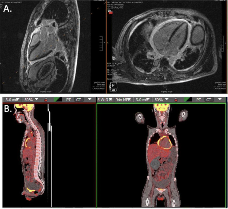
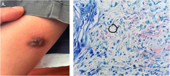
A 23-year-old previously healthy male presented to the hospital with symptoms of heart failure. He was diagnosed with pericarditis and found to have a reduced left ventricular ejection fraction of 25%. He was noted to have mediastinal lymphadenopathy. Pulmonary and abdominal sampling were non-diagnostic for infection, autoimmune disease, or malignancy. A QuantiFERON Gold returned positive. After a thorough travel history and detailed exam, the patient was diagnosed with disseminated tuberculosis after the discovery of a cutaneous gumma that was found to have acid-fast bacilli present on biopsy with Fite's stain. F-FDG PET CT and cardiac MRI were pursued given that pericardial and myocardial biopsy could not be safely performed due to the patient's hemodynamics. F-FDG PET CT and cardiac MRI did not demonstrate any myocardial pathology responsible for the left ventricular ejection fraction. This case highlights that pulmonary involvement is not necessary for disseminated TB, Fite's stain may be used to identify M. tuberculosis, and that cardiac MRI and F-FDG PET CT may be useful to delineate myocardial involvement in high-risk situations.

摘要

一位 23 岁既往健康的男性因心力衰竭症状到医院就诊。他被诊断为心包炎,并发现左心室射血分数降低至 25%。他被发现纵隔淋巴结肿大。肺部和腹部采样未发现感染、自身免疫性疾病或恶性肿瘤。QuantiFERON Gold 检测呈阳性。在详细的旅行史和检查后,患者被诊断为播散性结核,因为在活检中发现了皮肤树胶肿,Fite 染色显示有抗酸杆菌。由于患者的血液动力学不稳定,心包和心肌活检无法安全进行,因此进行了 F-FDG PET CT 和心脏 MRI。F-FDG PET CT 和心脏 MRI 均未显示任何导致左心室射血分数降低的心肌病变。本病例强调了肺部受累并非播散性结核所必需,Fite 染色可用于识别结核分枝杆菌,并且在高危情况下,心脏 MRI 和 F-FDG PET CT 可能有助于描绘心肌受累情况。

https://cdn.ncbi.nlm.nih.gov/pmc/blobs/6c1b/9360279/6a9882d14445/11606_2022_7619_Fig2_HTML.jpg
https://cdn.ncbi.nlm.nih.gov/pmc/blobs/6c1b/9360279/57f855650420/11606_2022_7619_Fig1_HTML.jpg
https://cdn.ncbi.nlm.nih.gov/pmc/blobs/6c1b/9360279/6a9882d14445/11606_2022_7619_Fig2_HTML.jpg
https://cdn.ncbi.nlm.nih.gov/pmc/blobs/6c1b/9360279/57f855650420/11606_2022_7619_Fig1_HTML.jpg
https://cdn.ncbi.nlm.nih.gov/pmc/blobs/6c1b/9360279/6a9882d14445/11606_2022_7619_Fig2_HTML.jpg

相似文献

1
Non-pulmonary Disseminated Tuberculosis Complicated by Constrictive Pericarditis and Cutaneous Gumma.非肺部播散性结核并发缩窄性心包炎和皮肤树胶肿。
J Gen Intern Med. 2022 Aug;37(10):2568-2572. doi: 10.1007/s11606-022-07619-w. Epub 2022 May 2.
2
Low-Flow, Low-Gradient Severe Aortic Stenosis in the Setting of Constrictive Pericarditis: Clinical Characteristics, Echocardiographic Features, and Outcomes.缩窄性心包炎背景下的低流量、低梯度重度主动脉瓣狭窄:临床特征、超声心动图特点及预后
Circ Cardiovasc Imaging. 2015 Jul;8(7):e002812. doi: 10.1161/CIRCIMAGING.114.002812.
3
Constrictive pericarditis in the setting of repeated chest trauma in a mixed martial arts fighter.在混合武术格斗者重复胸部创伤的情况下出现缩窄性心包炎。
BMC Cardiovasc Disord. 2021 Nov 22;21(1):561. doi: 10.1186/s12872-021-02378-8.
4
18F-FDG PET/CT Revealing Constrictive Pericarditis as the Only Manifestation of Malignant Mesothelioma.18F-FDG PET/CT 显示缩窄性心包炎为恶性间皮瘤的唯一表现。
Clin Nucl Med. 2019 Jan;44(1):55-56. doi: 10.1097/RLU.0000000000002349.
5
Gated F-18 FDG PET for assessment of left ventricular volumes and ejection fraction using QGS and 4D-MSPECT in patients with heart failure: a comparison with cardiac MRI.门控 F-18 FDG PET 用于评估心力衰竭患者左心室容积和射血分数:与心脏 MRI 的比较。
PLoS One. 2014 Jan 3;9(1):e80227. doi: 10.1371/journal.pone.0080227. eCollection 2014.
6
The role of F-fluorodeoxyglucose-positron emission tomography/computed tomography in the differential diagnosis of pericardial disease.氟代脱氧葡萄糖正电子发射断层扫描/计算机断层扫描在心包疾病鉴别诊断中的作用。
Sci Rep. 2020 Dec 9;10(1):21524. doi: 10.1038/s41598-020-78581-y.
7
Radionuclide angiography and magnetic resonance imaging: complementary non-invasive methods in the diagnosis of constrictive pericarditis.
Eur J Nucl Med. 1995 Nov;22(11):1292-8. doi: 10.1007/BF00801616.
8
[TUBERCULOUS CONSTRICTIVE PERICARDITIS DETECTED ON POSITRON EMISSION TOMOGRAPHY].[正电子发射断层扫描检测出的结核性缩窄性心包炎]
Kekkaku. 2016 Feb;91(2):65-8.
9
[Constrictive pericarditis illustrated by an unusual case report].[以一例罕见病例报告阐述缩窄性心包炎]
Tidsskr Nor Laegeforen. 1998 Nov 10;118(27):4217-21.
10
Breaking hearts and taking names: A case of sarcoidosis related effusive-constrictive pericarditis.伤心夺命:一例与结节病相关的心包渗出-缩窄性。
Respir Med. 2020 Mar;163:105879. doi: 10.1016/j.rmed.2020.105879. Epub 2020 Jan 23.

引用本文的文献

1
Unique pathological features and drug resistance patterns in cutaneous tuberculosis.皮肤结核的独特病理特征及耐药模式
Front Microbiol. 2025 May 30;16:1574051. doi: 10.3389/fmicb.2025.1574051. eCollection 2025.

本文引用的文献

1
An evolving presentation of cutaneous tuberculosis.皮肤结核的一种不断演变的表现形式。
Dermatol Online J. 2020 Aug 15;26(8):13030/qt55d3f43c.
2
Tuberculosis trends--United States, 2014.2014年美国结核病流行趋势
MMWR Morb Mortal Wkly Rep. 2015 Mar 20;64(10):265-9.
3
Tuberculous pericarditis presenting as massive haemorrhagic pericardial effusion.表现为大量出血性心包积液的结核性心包炎。
BMJ Case Rep. 2012 Jun 28;2012:bcr0320125967. doi: 10.1136/bcr.03.2012.5967.
4
Extrapulmonary tuberculosis: an overview.肺外结核病概述
Am Fam Physician. 2005 Nov 1;72(9):1761-8.
5
Experience with pericarditis at Groote Schuur Hospital, Cape Town: an analysis of one hundred and sixty cases studied over a six-year period.开普敦古特·舒尔医院心包炎的经验:对六年期间研究的160例病例的分析。
S Afr Med J. 1959 Sep 26;33:810-7.