Cho Sung-Min, Wilcox Christopher, Keller Steven, Acton Matthew, Rando Hannah, Etchill Eric, Giuliano Katherine, Bush Errol L, Sair Haris I, Pitts John, Kim Bo Soo, Whitman Glenn
Division of Cardiac Surgery, Department of Surgery, Johns Hopkins University School of Medicine, 600 N. Wolfe Street, Phipps 455, Baltimore, MD, 21287, USA.
Neuroscience Critical Care Division, Departments of Neurology, Neurosurgery, and Anesthesiology and Critical Care Medicine, Johns Hopkins University School of Medicine, Baltimore, MD, USA.
Crit Care. 2022 Apr 30;26(1):119. doi: 10.1186/s13054-022-03990-6.
To assess the safety and feasibility of imaging of the brain with a point-of-care (POC) magnetic resonance imaging (MRI) system in patients on extracorporeal membrane oxygenation (ECMO). Early detection of acute brain injury (ABI) is critical in improving survival for patients with ECMO support.
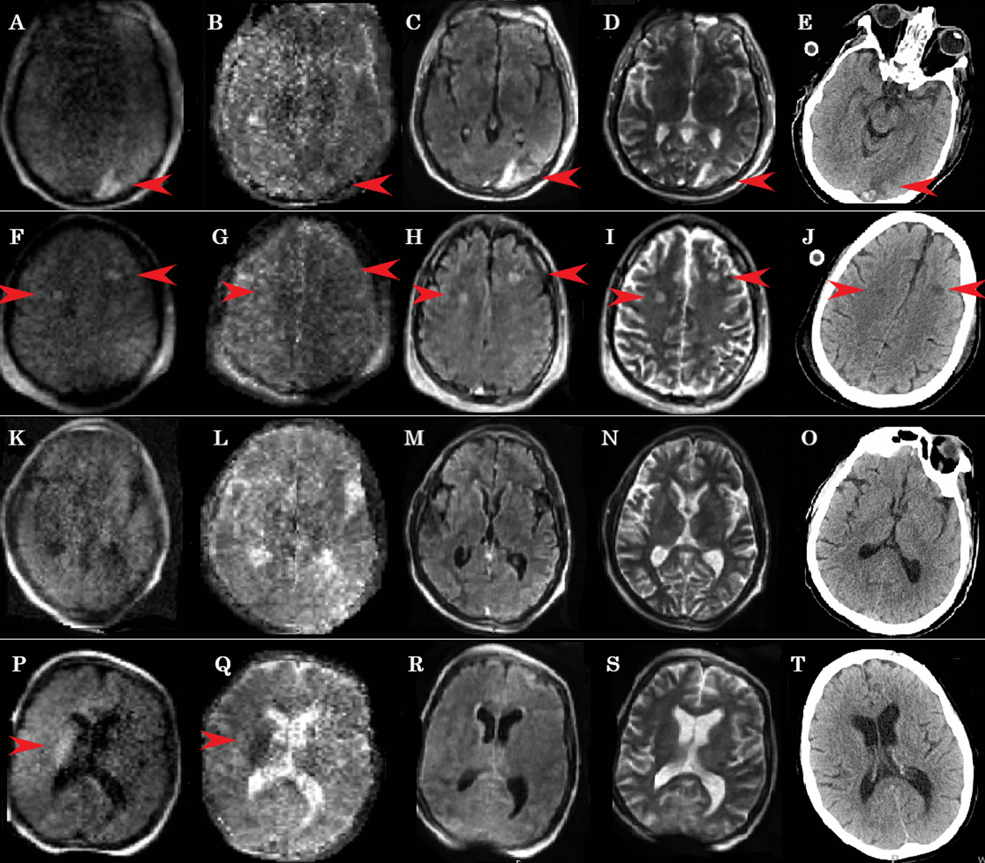
Patients from a single tertiary academic ECMO center who underwent head CT (HCT), followed by POC brain MRI examinations within 24 h following HCT while on ECMO. Primary outcomes were safety and feasibility, defined as completion of MRI examination without serious adverse events (SAEs). Secondary outcome was the quality of MR images in assessing ABIs.
We report 3 consecutive adult patients (median age 47 years; 67% male) with veno-arterial (n = 1) and veno-venous ECMO (n = 2) (VA- and VV-ECMO) support. All patients were imaged successfully without SAEs. Times to complete POC brain MRI examinations were 34, 40, and 43 min. Two patients had ECMO suction events, resolved with fluid and repositioning. Two patients were found to have an unsuspected acute stroke, well visualized with MRI.
Adult patients with VA- or VV-ECMO support can be safely imaged with low-field POC brain MRI in the intensive care unit, allowing for the assessment of presence and timing of ABI.
评估在接受体外膜肺氧合(ECMO)治疗的患者中,使用床旁(POC)磁共振成像(MRI)系统对脑部进行成像的安全性和可行性。早期发现急性脑损伤(ABI)对于提高接受ECMO支持的患者的生存率至关重要。
来自一个三级学术ECMO中心的患者,在接受头部CT(HCT)后,于接受ECMO治疗期间在HCT后24小时内接受床旁脑部MRI检查。主要结局是安全性和可行性,定义为在无严重不良事件(SAE)的情况下完成MRI检查。次要结局是MR图像在评估ABI方面的质量。
我们报告了3例连续的成年患者(中位年龄47岁;67%为男性),接受静脉-动脉(n = 1)和静脉-静脉ECMO(n = 2)(VA-和VV-ECMO)支持。所有患者均成功成像,无SAE。完成床旁脑部MRI检查的时间分别为34、40和43分钟。2例患者发生ECMO抽吸事件,通过补液和重新定位得以解决。2例患者被发现有意外的急性中风,MRI清晰显示。
在重症监护病房中,接受VA-或VV-ECMO支持的成年患者可以安全地使用低场床旁脑部MRI进行成像,从而能够评估ABI的存在和发生时间。