• 文献检索
  • 文档翻译
  • 深度研究
  • 学术资讯
  • Suppr Zotero 插件Zotero 插件
  • 邀请有礼
  • 套餐&价格
  • 历史记录
应用&插件
Suppr Zotero 插件Zotero 插件浏览器插件Mac 客户端Windows 客户端微信小程序
定价
高级版会员购买积分包购买API积分包
服务
文献检索文档翻译深度研究API 文档MCP 服务
关于我们
关于 Suppr公司介绍联系我们用户协议隐私条款
关注我们

Suppr 超能文献

核心技术专利:CN118964589B侵权必究
粤ICP备2023148730 号-1Suppr @ 2026

文献检索

告别复杂PubMed语法,用中文像聊天一样搜索,搜遍4000万医学文献。AI智能推荐,让科研检索更轻松。

立即免费搜索

文件翻译

保留排版,准确专业,支持PDF/Word/PPT等文件格式,支持 12+语言互译。

免费翻译文档

深度研究

AI帮你快速写综述,25分钟生成高质量综述,智能提取关键信息,辅助科研写作。

立即免费体验

一项由欧洲乳腺影像学学会开展的关于在临床实践中实施乳腺弥散加权成像的调查。

A survey by the European Society of Breast Imaging on the implementation of breast diffusion-weighted imaging in clinical practice.

机构信息

Department of Radiology, Breast Imaging Service, Memorial Sloan Kettering Cancer Center, 300 E 66th Street, New York, NY, 10065, USA.

Department of Epidemiology and Biostatistics, Memorial Sloan Kettering Cancer Center, 485 Lexington Ave, NY, New York, 10017, USA.

出版信息

Eur Radiol. 2022 Oct;32(10):6588-6597. doi: 10.1007/s00330-022-08833-0. Epub 2022 May 4.

DOI:10.1007/s00330-022-08833-0
PMID:35507050
原文链接:
https://pmc.ncbi.nlm.nih.gov/articles/PMC9064723/
Abstract

OBJECTIVES

To perform a survey among all European Society of Breast Imaging (EUSOBI) radiologist members to gather representative data regarding the clinical use of breast DWI.

METHODS

An online questionnaire was developed by two board-certified radiologists, reviewed by the EUSOBI board and committees, and finally distributed among EUSOBI active and associated (not based in Europe) radiologist members. The questionnaire included 20 questions pertaining to technical preferences (acquisition time, magnet strength, breast coils, number of b values), clinical indications, imaging evaluation, and reporting. Data were analyzed using descriptive statistics, the Chi-square test of independence, and Fisher's exact test.

RESULTS

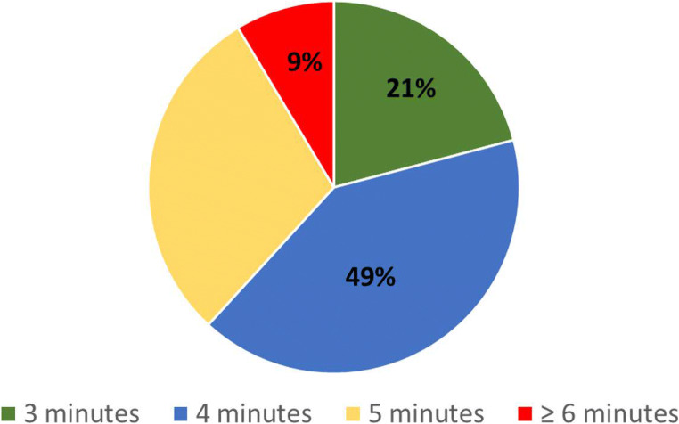
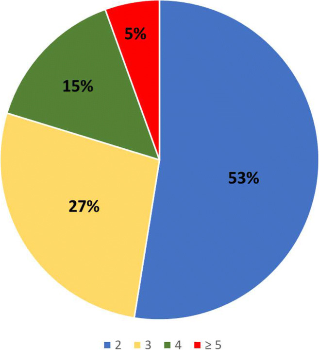
Of 1411 EUSOBI radiologist members, 275/1411 (19.5%) responded. Most (222/275, 81%) reported using DWI as part of their routine protocol. Common indications for DWI include lesion characterization (using an ADC threshold of 1.2-1.3 × 10 mm/s) and prediction of response to chemotherapy. Members most commonly acquire two separate b values (114/217, 53%), with b value = 800 s/mm being the preferred value for appraisal among those acquiring more than two b values (71/171, 42%). Most did not use synthetic b values (169/217, 78%). While most mention hindered diffusion in the MRI report (161/213, 76%), only 142/217 (57%) report ADC values.

CONCLUSION

The utilization of DWI in clinical practice among EUSOBI radiologists who responded to the survey is generally in line with international recommendations, with the main application being the differentiation of benign and malignant enhancing lesions, treatment response assessment, and prediction of response to chemotherapy. Report integration of qualitative and quantitative DWI data is not uniform.

KEY POINTS

• Clinical performance of breast DWI is in good agreement with the current recommendations of the EUSOBI International Breast DWI working group. • Breast DWI applications in clinical practice include the differentiation of benign and malignant enhancing, treatment response assessment, and prediction of response to chemotherapy. • Report integration of DWI results is not uniform.

摘要

目的

对所有欧洲乳腺影像学会(EUSOBI)放射科医师成员进行调查,以收集关于乳腺 DWI 临床应用的代表性数据。

方法

由两名经过董事会认证的放射科医师开发在线问卷,由 EUSOBI 董事会和委员会进行审查,最后分发给 EUSOBI 活跃成员和相关(不在欧洲的)成员。问卷包括 20 个问题,涉及技术偏好(采集时间、磁场强度、乳腺线圈、b 值数量)、临床指征、成像评估和报告。使用描述性统计、独立性卡方检验和 Fisher 精确检验进行数据分析。

结果

在 1411 名 EUSOBI 放射科医师成员中,有 275/1411(19.5%)做出回应。大多数(222/275,81%)报告将 DWI 作为其常规方案的一部分。DWI 的常见指征包括病变特征(使用 1.2-1.3×10 mm/s 的 ADC 阈值)和预测化疗反应。成员最常获取两个单独的 b 值(114/217,53%),在获取两个以上 b 值的情况下,b 值=800 s/mm 是最受欢迎的评估值(71/171,42%)。大多数人不使用合成 b 值(169/217,78%)。虽然大多数人在 MRI 报告中提到扩散受限(161/213,76%),但只有 142/217(57%)报告 ADC 值。

结论

在回应调查的 EUSOBI 放射科医师中,DWI 在临床实践中的应用通常符合国际建议,主要应用是区分良性和恶性强化病变、治疗反应评估和预测化疗反应。定性和定量 DWI 数据的报告整合并不统一。

重点

  1. 乳腺 DWI 的临床性能与 EUSOBI 国际乳腺 DWI 工作组的现行建议基本一致。

  2. 乳腺 DWI 在临床实践中的应用包括良性和恶性强化病变的鉴别、治疗反应评估和预测化疗反应。

  3. DWI 结果的报告整合并不统一。

https://cdn.ncbi.nlm.nih.gov/pmc/blobs/d5b4/9064723/bc75f8ae74f5/330_2022_8833_Fig2_HTML.jpg
https://cdn.ncbi.nlm.nih.gov/pmc/blobs/d5b4/9064723/1bbbced969b5/330_2022_8833_Fig1_HTML.jpg
https://cdn.ncbi.nlm.nih.gov/pmc/blobs/d5b4/9064723/bc75f8ae74f5/330_2022_8833_Fig2_HTML.jpg
https://cdn.ncbi.nlm.nih.gov/pmc/blobs/d5b4/9064723/1bbbced969b5/330_2022_8833_Fig1_HTML.jpg
https://cdn.ncbi.nlm.nih.gov/pmc/blobs/d5b4/9064723/bc75f8ae74f5/330_2022_8833_Fig2_HTML.jpg

相似文献

1
A survey by the European Society of Breast Imaging on the implementation of breast diffusion-weighted imaging in clinical practice.一项由欧洲乳腺影像学学会开展的关于在临床实践中实施乳腺弥散加权成像的调查。
Eur Radiol. 2022 Oct;32(10):6588-6597. doi: 10.1007/s00330-022-08833-0. Epub 2022 May 4.
2
Diffusion-weighted imaging of the breast-a consensus and mission statement from the EUSOBI International Breast Diffusion-Weighted Imaging working group.乳腺弥散加权成像的共识和任务声明:来自 EUSOBI 国际乳腺弥散加权成像工作组。
Eur Radiol. 2020 Mar;30(3):1436-1450. doi: 10.1007/s00330-019-06510-3. Epub 2019 Nov 30.
3
A survey by the European Society of Breast Imaging on the utilisation of breast MRI in clinical practice.欧洲乳腺影像学会关于乳腺 MRI 在临床实践中应用的调查。
Eur Radiol. 2018 May;28(5):1909-1918. doi: 10.1007/s00330-017-5121-4. Epub 2017 Nov 22.
4
Added value of the EUSOBI diffusion levels in breast MRI.EUSOBI 弥散水平在乳腺 MRI 中的附加价值。
Eur Radiol. 2024 May;34(5):3352-3363. doi: 10.1007/s00330-023-10418-4. Epub 2023 Nov 7.
5
Advanced breast diffusion-weighted imaging: what are the next steps? A proposal from the EUSOBI International Breast Diffusion-weighted Imaging working group.晚期乳腺扩散加权成像:下一步该怎么做?欧洲乳腺影像学会国际乳腺扩散加权成像工作组的提议
Eur Radiol. 2025 Apr;35(4):2130-2140. doi: 10.1007/s00330-024-11010-0. Epub 2024 Oct 8.
6
Current use and future perspectives of contrast-enhanced mammography (CEM): a survey by the European Society of Breast Imaging (EUSOBI).当前对比增强乳腺摄影(CEM)的使用情况和未来展望:欧洲乳腺影像学会(EUSOBI)的调查。
Eur Radiol. 2024 Aug;34(8):5439-5450. doi: 10.1007/s00330-023-10574-7. Epub 2024 Jan 16.
7
Can DWI provide additional value to Kaiser score in evaluation of breast lesions.DWI 是否能为 Kaiser 评分在评估乳腺病变中的应用提供额外价值。
Eur Radiol. 2022 Sep;32(9):5964-5973. doi: 10.1007/s00330-022-08674-x. Epub 2022 Mar 31.
8
MRI-only lesions: application of diffusion-weighted imaging obviates unnecessary MR-guided breast biopsies.仅 MRI 病灶:弥散加权成像的应用可避免不必要的 MR 引导下乳腺活检。
Eur Radiol. 2014 Jun;24(6):1204-10. doi: 10.1007/s00330-014-3153-6. Epub 2014 Apr 5.
9
Additional value of diffusion-weighted imaging to evaluate multifocal and multicentric breast cancer detected using pre-operative breast MRI.术前乳腺 MRI 检出多灶性和多中心性乳腺癌中弥散加权成像的附加价值。
Eur Radiol. 2017 Nov;27(11):4819-4827. doi: 10.1007/s00330-017-4898-5. Epub 2017 Jun 7.
10
Assessment of breast mass morphology with diffusion-weighted MRI: Beyond apparent diffusion coefficient.评估乳腺肿块形态学的弥散加权 MRI:超越表观扩散系数。
J Magn Reson Imaging. 2018 Dec;48(6):1668-1677. doi: 10.1002/jmri.26175. Epub 2018 May 7.

引用本文的文献

1
Can a low b-value diffusion-weighted imaging sequence replace the standard T2-weighted fluid-sensitive sequence in breast MRI? A single-center prospective study.在乳腺磁共振成像中,低b值扩散加权成像序列能否取代标准的T2加权液体敏感序列?一项单中心前瞻性研究。
Eur Radiol. 2025 May 7. doi: 10.1007/s00330-025-11624-y.
2
Reduction of biopsy rate in BI-RADS4 breast lesions: potential of an abbreviated advanced DWI protocol.降低乳腺影像报告和数据系统(BI-RADS)4类乳腺病变的活检率:简化的高级扩散加权成像(DWI)方案的潜力
Eur Radiol. 2025 Apr 24. doi: 10.1007/s00330-025-11604-2.
3
Comparative analysis of apparent diffusion coefficient (ADC) metrics for the differential diagnosis of breast mass lesions.

本文引用的文献

1
Diffusion-Weighted Magnetic Resonance Imaging for Breast Cancer Screening in High-Risk Women: Design and Imaging Protocol of a Prospective Multicenter Study in Korea.用于高危女性乳腺癌筛查的扩散加权磁共振成像:韩国一项前瞻性多中心研究的设计与成像方案
J Breast Cancer. 2021 Apr;24(2):218-228. doi: 10.4048/jbc.2021.24.e19.
2
Diffusion-weighted Imaging Allows for Downgrading MR BI-RADS 4 Lesions in Contrast-enhanced MRI of the Breast to Avoid Unnecessary Biopsy.弥散加权成像可将乳腺增强 MRI 中 BI-RADS 4 级病变降级,避免不必要的活检。
Clin Cancer Res. 2021 Apr 1;27(7):1941-1948. doi: 10.1158/1078-0432.CCR-20-3037. Epub 2021 Jan 14.
3
表观扩散系数(ADC)指标在乳腺肿块病变鉴别诊断中的对比分析
BMC Med Imaging. 2025 Apr 11;25(1):117. doi: 10.1186/s12880-025-01654-9.
4
Retrospective BReast Intravoxel Incoherent Motion Multisite (BRIMM) multisoftware study.回顾性乳腺体素内不相干运动多中心(BRIMM)多软件研究。
Front Oncol. 2025 Feb 24;15:1524634. doi: 10.3389/fonc.2025.1524634. eCollection 2025.
5
External validation of multiparametric magnetic resonance imaging-based decision rules for characterizing breast lesions and comparison to Kaiser score and breast imaging reporting and data system (BI-RADS) category.基于多参数磁共振成像的乳腺病变特征描述决策规则的外部验证以及与凯泽评分和乳腺影像报告和数据系统(BI-RADS)分类的比较
Quant Imaging Med Surg. 2025 Jan 2;15(1):648-661. doi: 10.21037/qims-23-1783. Epub 2024 Dec 30.
6
Diffusion weighted imaging for improving the diagnostic performance of screening breast MRI: impact of apparent diffusion coefficient quantitation methods and cutoffs.扩散加权成像用于提高乳腺磁共振成像筛查的诊断性能:表观扩散系数定量方法及临界值的影响
Front Oncol. 2024 Dec 20;14:1437506. doi: 10.3389/fonc.2024.1437506. eCollection 2024.
7
Diffusion-Weighted MRI for the Assessment of Molecular Prognostic Biomarkers in Breast Cancer.扩散加权磁共振成像在乳腺癌分子预后生物标志物评估中的应用。
Korean J Radiol. 2024 Jul;25(7):623-633. doi: 10.3348/kjr.2023.1188.
8
Diffusion-Weighted Imaging for Skin Pathologies of the Breast-A Feasibility Study.乳腺皮肤病变的扩散加权成像——一项可行性研究
Diagnostics (Basel). 2024 Apr 29;14(9):934. doi: 10.3390/diagnostics14090934.
9
Added value of the EUSOBI diffusion levels in breast MRI.EUSOBI 弥散水平在乳腺 MRI 中的附加价值。
Eur Radiol. 2024 May;34(5):3352-3363. doi: 10.1007/s00330-023-10418-4. Epub 2023 Nov 7.
10
Quantitative apparent diffusion coefficient metrics for MRI-only suspicious breast lesions: any added clinical value?仅基于MRI的可疑乳腺病变的定量表观扩散系数指标:有额外的临床价值吗?
Quant Imaging Med Surg. 2023 Oct 1;13(10):7092-7104. doi: 10.21037/qims-23-331. Epub 2023 Sep 12.
Breast arterial calcifications as a biomarker of cardiovascular risk: radiologists' awareness, reporting, and action. A survey among the EUSOBI members.
乳腺动脉钙化作为心血管风险的生物标志物:放射科医生的认识、报告和行动。一项对 EUSOBI 成员的调查。
Eur Radiol. 2021 Feb;31(2):958-966. doi: 10.1007/s00330-020-07136-6. Epub 2020 Aug 27.
4
Diagnostic value of diffusion-weighted imaging with synthetic b-values in breast tumors: comparison with dynamic contrast-enhanced and multiparametric MRI.扩散加权成像合成 b 值对乳腺肿瘤的诊断价值:与动态对比增强和多参数 MRI 的比较。
Eur Radiol. 2021 Jan;31(1):356-367. doi: 10.1007/s00330-020-07094-z. Epub 2020 Aug 11.
5
Feasibility Study of Synthetic Diffusion-Weighted MRI in Patients with Breast Cancer in Comparison with Conventional Diffusion-Weighted MRI.合成扩散加权 MRI 与常规扩散加权 MRI 对比在乳腺癌患者中的可行性研究。
Korean J Radiol. 2020 Sep;21(9):1036-1044. doi: 10.3348/kjr.2019.0568.
6
Diffusion-weighted imaging of the breast-a consensus and mission statement from the EUSOBI International Breast Diffusion-Weighted Imaging working group.乳腺弥散加权成像的共识和任务声明:来自 EUSOBI 国际乳腺弥散加权成像工作组。
Eur Radiol. 2020 Mar;30(3):1436-1450. doi: 10.1007/s00330-019-06510-3. Epub 2019 Nov 30.
7
Diffusion-weighted MRI for Unenhanced Breast Cancer Screening.弥散加权 MRI 在乳腺癌筛查中的应用。
Radiology. 2019 Dec;293(3):504-520. doi: 10.1148/radiol.2019182789. Epub 2019 Oct 8.
8
Diffusion-Weighted MRI of Breast Cancer: Improved Lesion Visibility and Image Quality Using Synthetic b-Values.乳腺癌的弥散加权 MRI:使用合成 b 值提高病变可见度和图像质量。
J Magn Reson Imaging. 2019 Dec;50(6):1754-1761. doi: 10.1002/jmri.26809. Epub 2019 May 28.
9
A Meta-analysis of the Diagnostic Performance of Diffusion MRI for Breast Lesion Characterization.磁共振弥散成像对乳腺病变特征诊断性能的 Meta 分析。
Radiology. 2019 Jun;291(3):632-641. doi: 10.1148/radiol.2019182510. Epub 2019 Apr 23.
10
Contrast-enhanced MRI for breast cancer screening.对比增强磁共振成像在乳腺癌筛查中的应用。
J Magn Reson Imaging. 2019 Aug;50(2):377-390. doi: 10.1002/jmri.26654. Epub 2019 Jan 18.