• 文献检索
  • 文档翻译
  • 深度研究
  • 学术资讯
  • Suppr Zotero 插件Zotero 插件
  • 邀请有礼
  • 套餐&价格
  • 历史记录
应用&插件
Suppr Zotero 插件Zotero 插件浏览器插件Mac 客户端Windows 客户端微信小程序
定价
高级版会员购买积分包购买API积分包
服务
文献检索文档翻译深度研究API 文档MCP 服务
关于我们
关于 Suppr公司介绍联系我们用户协议隐私条款
关注我们

Suppr 超能文献

核心技术专利:CN118964589B侵权必究
粤ICP备2023148730 号-1Suppr @ 2026

文献检索

告别复杂PubMed语法,用中文像聊天一样搜索,搜遍4000万医学文献。AI智能推荐,让科研检索更轻松。

立即免费搜索

文件翻译

保留排版,准确专业,支持PDF/Word/PPT等文件格式,支持 12+语言互译。

免费翻译文档

深度研究

AI帮你快速写综述,25分钟生成高质量综述,智能提取关键信息,辅助科研写作。

立即免费体验

灵活多变:分泌肾素细胞的可塑性。

Flexible and multifaceted: the plasticity of renin-expressing cells.

机构信息

Institute of Physiology, University of Regensburg, Universitätsstraβe 31, D-93053 , Regensburg, Germany.

出版信息

Pflugers Arch. 2022 Aug;474(8):799-812. doi: 10.1007/s00424-022-02694-8. Epub 2022 May 5.

DOI:10.1007/s00424-022-02694-8
PMID:35511367
原文链接:https://pmc.ncbi.nlm.nih.gov/articles/PMC9338909/
Abstract

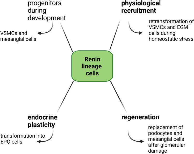
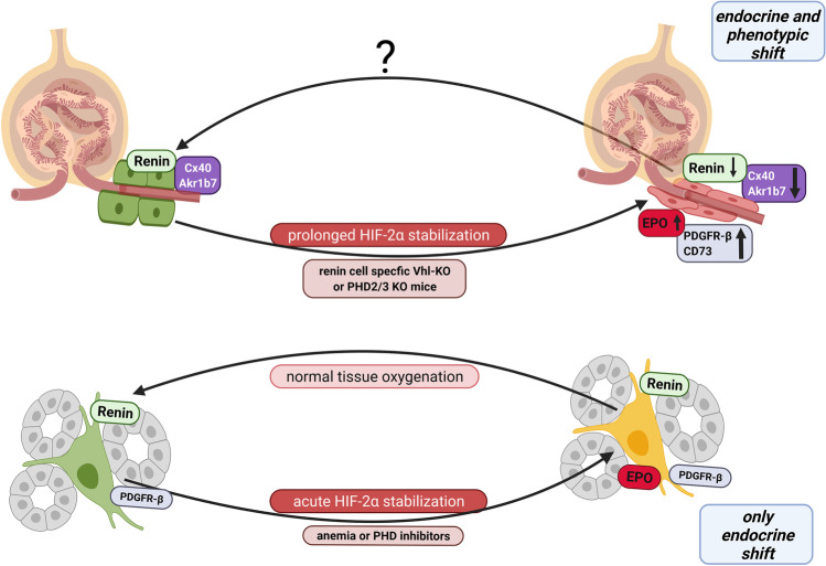
The protease renin, the key enzyme of the renin-angiotensin-aldosterone system, is mainly produced and secreted by juxtaglomerular cells in the kidney, which are located in the walls of the afferent arterioles at their entrance into the glomeruli. When the body's demand for renin rises, the renin production capacity of the kidneys commonly increases by induction of renin expression in vascular smooth muscle cells and in extraglomerular mesangial cells. These cells undergo a reversible metaplastic cellular transformation in order to produce renin. Juxtaglomerular cells of the renin lineage have also been described to migrate into the glomerulus and differentiate into podocytes, epithelial cells or mesangial cells to restore damaged cells in states of glomerular disease. More recently, it could be shown that renin cells can also undergo an endocrine and metaplastic switch to erythropoietin-producing cells. This review aims to describe the high degree of plasticity of renin-producing cells of the kidneys and to analyze the underlying mechanisms.

摘要

肾素酶,肾素-血管紧张素-醛固酮系统的关键酶,主要由肾脏的球旁细胞产生和分泌,这些细胞位于入球小动脉的壁内,就在它们进入肾小球的地方。当身体对肾素的需求增加时,肾脏通常通过诱导血管平滑肌细胞和肾小球系膜细胞中的肾素表达来增加肾素的产生能力。这些细胞经历可逆的细胞转化,以产生肾素。肾素谱系的球旁细胞也已被描述为迁移到肾小球并分化为足细胞、上皮细胞或系膜细胞,以在肾小球疾病状态下修复受损的细胞。最近,有人证明肾素细胞也可以发生内分泌和细胞转化为产生促红细胞生成素的细胞。这篇综述旨在描述肾脏产生肾素的细胞的高度可塑性,并分析其潜在机制。

https://cdn.ncbi.nlm.nih.gov/pmc/blobs/fab6/9338909/e47d2b74366d/424_2022_2694_Fig4_HTML.jpg
https://cdn.ncbi.nlm.nih.gov/pmc/blobs/fab6/9338909/0454d78dc0cc/424_2022_2694_Fig1_HTML.jpg
https://cdn.ncbi.nlm.nih.gov/pmc/blobs/fab6/9338909/5ed0bf09d6c7/424_2022_2694_Fig2_HTML.jpg
https://cdn.ncbi.nlm.nih.gov/pmc/blobs/fab6/9338909/8ba173ea8c87/424_2022_2694_Fig3_HTML.jpg
https://cdn.ncbi.nlm.nih.gov/pmc/blobs/fab6/9338909/e47d2b74366d/424_2022_2694_Fig4_HTML.jpg

相似文献

1
Flexible and multifaceted: the plasticity of renin-expressing cells.灵活多变:分泌肾素细胞的可塑性。
Pflugers Arch. 2022 Aug;474(8):799-812. doi: 10.1007/s00424-022-02694-8. Epub 2022 May 5.
2
Connexin Signaling in the Juxtaglomerular Apparatus (JGA) of Developing, Postnatal Healthy and Nephrotic Human Kidneys.发育、出生后健康和肾病人类肾脏中的连接蛋白信号转导在肾小球旁器(JGA)中。
Int J Mol Sci. 2020 Nov 6;21(21):8349. doi: 10.3390/ijms21218349.
3
High-level connexin expression in the human juxtaglomerular apparatus.人肾小球旁器中的高表达连接蛋白。
Nephron Physiol. 2010;116(1):p1-8. doi: 10.1159/000315658. Epub 2010 Jun 8.
4
Cells of renin lineage are adult pluripotent progenitors in experimental glomerular disease.肾素谱系细胞是实验性肾小球疾病中的成年多能祖细胞。
Am J Physiol Renal Physiol. 2015 Aug 15;309(4):F341-58. doi: 10.1152/ajprenal.00438.2014. Epub 2015 Jun 10.
5
Progenitor Renin Lineage Cells are not involved in the regeneration of glomerular endothelial cells during experimental renal thrombotic microangiopathy.祖细胞肾素谱系细胞不参与实验性肾血栓性微血管病期间肾小球内皮细胞的再生。
PLoS One. 2018 May 17;13(5):e0196752. doi: 10.1371/journal.pone.0196752. eCollection 2018.
6
Piezo2 expression and its alteration by mechanical forces in mouse mesangial cells and renin-producing cells.Piezo2 在小鼠系膜细胞和肾素产生细胞中的表达及其机械力改变。
Sci Rep. 2022 Mar 10;12(1):4197. doi: 10.1038/s41598-022-07987-7.
7
Sox6 as a new modulator of renin expression in the kidney.Sox6 作为肾脏中肾素表达的新调节因子。
Am J Physiol Renal Physiol. 2020 Feb 1;318(2):F285-F297. doi: 10.1152/ajprenal.00095.2019. Epub 2019 Nov 25.
8
Lack of connexin 40 causes displacement of renin-producing cells from afferent arterioles to the extraglomerular mesangium.连接蛋白40的缺失导致产肾素细胞从入球小动脉移位至球外系膜。
J Am Soc Nephrol. 2007 Apr;18(4):1103-11. doi: 10.1681/ASN.2006090953. Epub 2007 Feb 28.
9
Adult renal mesenchymal stem cell-like cells contribute to juxtaglomerular cell recruitment.成人肾间质干细胞样细胞有助于球旁细胞的募集。
J Am Soc Nephrol. 2013 Jul;24(8):1263-73. doi: 10.1681/ASN.2012060596. Epub 2013 Jun 6.
10
Connexin 40 is dispensable for vascular renin cell recruitment but is indispensable for vascular baroreceptor control of renin secretion.连接蛋白40对血管肾素细胞募集并非必需,但对肾素分泌的血管压力感受器控制却是不可或缺的。
Pflugers Arch. 2015 Aug;467(8):1825-34. doi: 10.1007/s00424-014-1615-y. Epub 2014 Sep 23.

引用本文的文献

1
Direct Vascular Effects of Angiotensin II (A Systematic Short Review).血管紧张素II的直接血管效应(系统简要综述)
Int J Mol Sci. 2024 Dec 26;26(1):113. doi: 10.3390/ijms26010113.
2
A Possible Link between Cell Plasticity and Renin Expression in the Collecting Duct: A Narrative Review.收集管中细胞可塑性与肾素表达之间的可能联系:叙事性综述。
Int J Mol Sci. 2024 Sep 3;25(17):9549. doi: 10.3390/ijms25179549.
3
Hold the salt for kidney regeneration.少吃盐有助于肾脏再生。

本文引用的文献

1
The Transcription Factor Sox6 Controls Renin Expression during Renal Artery Stenosis.转录因子 Sox6 在肾动脉狭窄期间控制肾素表达。
Kidney360. 2021 Mar 26;2(5):842-856. doi: 10.34067/KID.0002792020. eCollection 2021 May 27.
2
Intact prostaglandin signaling through EP2 and EP4 receptors in stromal progenitor cells is required for normal development of the renal cortex in mice.在小鼠中,基质祖细胞中 EP2 和 EP4 受体的完整前列腺素信号传导对于肾皮质的正常发育是必需的。
Am J Physiol Renal Physiol. 2022 Mar 1;322(3):F295-F307. doi: 10.1152/ajprenal.00414.2021. Epub 2022 Jan 17.
3
Prolyl-4-hydroxylases 2 and 3 control erythropoietin production in renin-expressing cells of mouse kidneys.
J Clin Invest. 2024 Jun 3;134(11):e181397. doi: 10.1172/JCI181397.
4
Salt and Aldosterone - Reciprocal and Combined Effects in Preclinical Models and Humans.盐和醛固酮——临床前模型和人类中的相互作用和联合效应。
Horm Metab Res. 2024 Jan;56(1):99-106. doi: 10.1055/a-2172-7228. Epub 2023 Sep 8.
5
The kidneys matter.肾脏至关重要。
Pflugers Arch. 2022 Aug;474(8):755-757. doi: 10.1007/s00424-022-02737-0.
脯氨酰-4-羟化酶2和3控制小鼠肾脏中表达肾素细胞的促红细胞生成素产生。
J Physiol. 2022 Feb;600(3):671-694. doi: 10.1113/JP282615. Epub 2021 Dec 27.
4
Inhibition of the renin-angiotensin system causes concentric hypertrophy of renal arterioles in mice and humans.肾素-血管紧张素系统的抑制会导致小鼠和人类肾小动脉的向心性肥大。
JCI Insight. 2021 Dec 22;6(24):e154337. doi: 10.1172/jci.insight.154337.
5
Patterns of differentiation of renin lineage cells during nephrogenesis.肾素谱系细胞在肾发生过程中的分化模式。
Am J Physiol Renal Physiol. 2021 Sep 1;321(3):F378-F388. doi: 10.1152/ajprenal.00151.2021. Epub 2021 Aug 2.
6
Renin Cells, the Kidney, and Hypertension.肾素细胞、肾脏和高血压。
Circ Res. 2021 Apr 2;128(7):887-907. doi: 10.1161/CIRCRESAHA.121.318064. Epub 2021 Apr 1.
7
Hypoxia-inducible factor-prolyl hydroxylase inhibitors in the treatment of anemia of chronic kidney disease.缺氧诱导因子脯氨酰羟化酶抑制剂在治疗慢性肾脏病贫血中的应用
Kidney Int Suppl (2011). 2021 Apr;11(1):8-25. doi: 10.1016/j.kisu.2020.12.002. Epub 2021 Mar 18.
8
Localization of angiotensin II type 1 receptor gene expression in rodent and human kidneys.血管紧张素 II 型 1 受体基因在啮齿动物和人类肾脏中的表达定位。
Am J Physiol Renal Physiol. 2021 Apr 1;320(4):F644-F653. doi: 10.1152/ajprenal.00550.2020. Epub 2021 Feb 22.
9
Renal interstitial fibroblasts coproduce erythropoietin and renin under anaemic conditions.在贫血条件下,肾间质成纤维细胞共同产生促红细胞生成素和肾素。
EBioMedicine. 2021 Feb;64:103209. doi: 10.1016/j.ebiom.2021.103209. Epub 2021 Jan 25.
10
Transcription Factor RBPJ as a Molecular Switch in Regulating the Notch Response.转录因子 RBPJ 作为调节 Notch 反应的分子开关。
Adv Exp Med Biol. 2021;1287:9-30. doi: 10.1007/978-3-030-55031-8_2.