García Feijoo Pablo, Carceller Fernando, Isla Guerrero Alberto, Sáez-Alegre Miguel, Gandía González Maria Luisa
Department of Neurosurgery, La Paz University Hospital, Madrid, Spain.
Front Surg. 2022 Apr 18;9:884675. doi: 10.3389/fsurg.2022.884675. eCollection 2022.
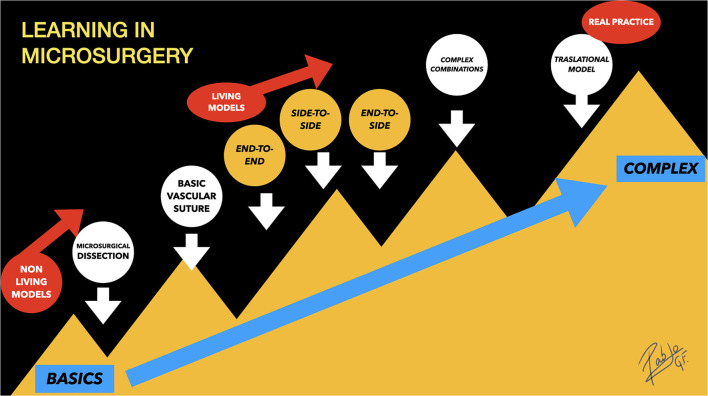
Nowadays, due to the decline in the number of microsurgical clippings for cerebral aneurysms and revascularization procedures, young neurosurgeons have fewer opportunities to participate and train on this type of surgery. Vascular neurosurgery is a demanding subspecialty that requires skills that can only be acquired with technical experience. This background pushes the new generations to be ready for such challenging cases by training hard on different available models, such as synthetic tubes, chicken wings, or placenta vessels. Although many training models for vascular neurosurgery have been described worldwide, one of the best is the rodent vessels model. It offers pulsation, coagulation, and real blood flow conditions in a physiologic atmosphere that mimics perfectly the intracranial human vessels environment, especially in terms of size. However, the current differences in governmental different regulations about the use of living animals in medical experimentation and the social awareness, as well as the lack of financial support, cause more difficulties for neurosurgeons to start with that kind of training. In this review, we describe the tools and techniques as basic steps for vascular microsurgery training by using rodent models, that provide an accurate copy of brain vessels environment under stable conditions. The initial three classical known microanastomoses for neurosurgeons are end-to-end, end-to-side, and side-to-side, but in literature, there have been described other more complex exercises for training and investigation, such as aneurysm models. Although there is still little data available, we aim to summarize and discuss aneurysm's training models and reviewed the current literature on the subject and its applications, including a detailed description of the techniques.
如今,由于用于脑动脉瘤的显微外科夹闭术和血管重建手术的数量减少,年轻神经外科医生参与此类手术并接受培训的机会也随之减少。血管神经外科是一个要求很高的亚专业,需要通过技术经验才能获得相关技能。这种背景促使新一代通过在不同的可用模型上刻苦训练,如合成管、鸡翅或胎盘血管,为应对此类具有挑战性的病例做好准备。尽管全球已经描述了许多用于血管神经外科的训练模型,但其中最好的之一是啮齿动物血管模型。它在生理环境中提供搏动、凝血和真实血流情况,完美模拟颅内人体血管环境,尤其是在尺寸方面。然而,目前政府关于在医学实验中使用活体动物的不同法规差异、社会意识以及缺乏资金支持,给神经外科医生开展此类训练带来了更多困难。在本综述中,我们描述了使用啮齿动物模型进行血管显微外科训练的工具和技术,这些技术是血管显微外科训练的基本步骤,能在稳定条件下精确复制脑血管环境。神经外科医生最初熟知的三种经典显微吻合方式是端端吻合、端侧吻合和侧侧吻合,但文献中还描述了其他更复杂的训练和研究练习,如动脉瘤模型。尽管目前可用数据仍然很少,但我们旨在总结和讨论动脉瘤训练模型,回顾该主题的当前文献及其应用,包括技术的详细描述。