Labcorp Drug Development, Morrisville, North Carolina, USA.
Labcorp of America, Burlington, North Carolina, USA.
J Clin Pathol. 2023 Sep;76(9):591-598. doi: 10.1136/jclinpath-2022-208254. Epub 2022 May 9.
A robust immunohistochemistry (IHC) assay was developed to detect lymphocyte-activation gene 3 (LAG-3) expression by immune cells (ICs) in tumour tissues. LAG-3 is an immuno-oncology target with demonstrable clinical benefit, and there is a need for a standardised, well-characterised assay to measure its expression. This study aims to describe LAG-3 scoring criteria and present the specificity, sensitivity, analytical precision and reproducibility of this assay.
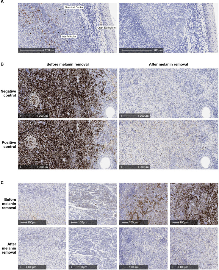
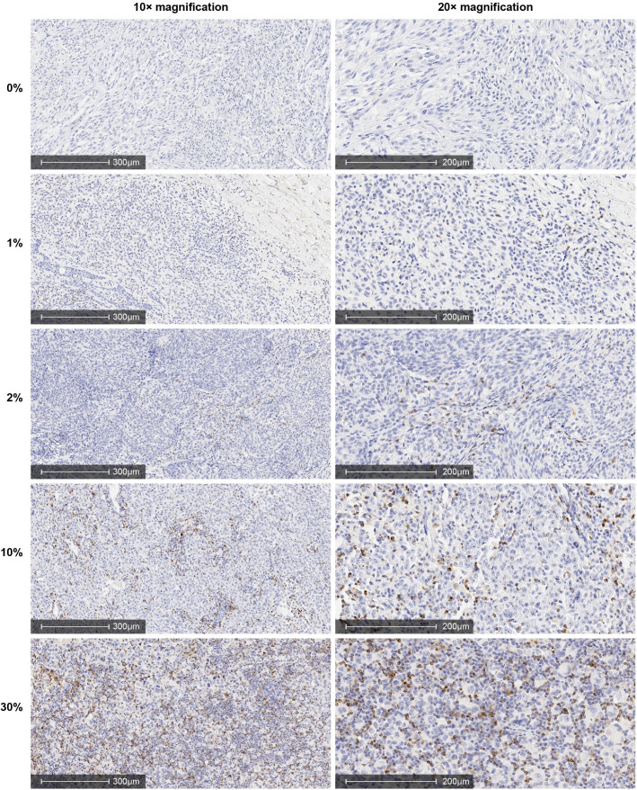
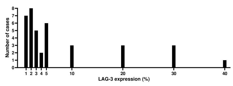
The specificity of the assay was investigated by antigen competition and with knockout cell lines. A melanin pigment removal procedure was implemented to prevent melanin interference in IHC interpretation. Formalin-fixed paraffin-embedded (FFPE) human melanoma samples with a range of LAG-3 expression levels were used to assess the sensitivity and analytical precision of the assay with a ≥1% cut-off to determine LAG-3 positivity. Interobserver and intraobserver reproducibility were evaluated with 60 samples in intralaboratory studies and 70 samples in interlaboratory studies.
The LAG-3 IHC method demonstrated performance suitable for analysis of LAG-3 IC expression in clinical melanoma samples. The pretreatment step effectively removed melanin pigment that could interfere with interpretation. LAG-3 antigen competition and analysis of knockout cell lines indicated that the 17B4 antibody clone binds specifically to LAG-3. The intrarun repeatability, interday, interinstrument, interoperator and inter-reagent lot reproducibility demonstrated a high scoring concordance (>95%). The interobserver and intraobserver reproducibility and overall interlaboratory and intralaboratory reproducibility also showed high scoring concordance (>90%).
We have demonstrated that the assay reliably assesses LAG-3 expression in FFPE human melanoma samples by IHC.
开发了一种稳健的免疫组织化学(IHC)检测方法,用于检测肿瘤组织中免疫细胞(ICs)的淋巴细胞激活基因 3(LAG-3)表达。LAG-3 是一种具有明显临床获益的免疫肿瘤学靶点,因此需要一种标准化、特征明确的检测方法来测量其表达。本研究旨在描述 LAG-3 评分标准,并介绍该检测方法的特异性、敏感性、分析精密度和可重复性。
通过抗原竞争和 基因敲除细胞系来研究该检测方法的特异性。采用黑色素去除程序来防止黑色素对 IHC 解读的干扰。使用具有不同 LAG-3 表达水平的福尔马林固定石蜡包埋(FFPE)人黑色素瘤样本评估该检测方法的敏感性和分析精密度,以 ≥1%的截断值确定 LAG-3 阳性。在实验室内部研究中评估了 60 个样本的观察者间和观察者内的可重复性,在实验室间研究中评估了 70 个样本的可重复性。
LAG-3 IHC 方法在分析临床黑色素瘤样本中的 LAG-3 IC 表达方面表现出了适合的性能。预处理步骤可有效去除可能干扰解读的黑色素。LAG-3 抗原竞争和 基因敲除细胞系的分析表明,17B4 抗体克隆特异性结合 LAG-3。日内、日间、仪器间、操作者间和试剂批次间的重复性试验显示,评分一致性高(>95%)。观察者间、观察者内的可重复性以及总体实验室间和实验室内的可重复性也显示出了高评分一致性(>90%)。
我们已经证明,该检测方法通过 IHC 可可靠地评估 FFPE 人黑色素瘤样本中的 LAG-3 表达。