• 文献检索
  • 文档翻译
  • 深度研究
  • 学术资讯
  • Suppr Zotero 插件Zotero 插件
  • 邀请有礼
  • 套餐&价格
  • 历史记录
应用&插件
Suppr Zotero 插件Zotero 插件浏览器插件Mac 客户端Windows 客户端微信小程序
定价
高级版会员购买积分包购买API积分包
服务
文献检索文档翻译深度研究API 文档MCP 服务
关于我们
关于 Suppr公司介绍联系我们用户协议隐私条款
关注我们

Suppr 超能文献

核心技术专利:CN118964589B侵权必究
粤ICP备2023148730 号-1Suppr @ 2026

文献检索

告别复杂PubMed语法,用中文像聊天一样搜索,搜遍4000万医学文献。AI智能推荐,让科研检索更轻松。

立即免费搜索

文件翻译

保留排版,准确专业,支持PDF/Word/PPT等文件格式,支持 12+语言互译。

免费翻译文档

深度研究

AI帮你快速写综述,25分钟生成高质量综述,智能提取关键信息,辅助科研写作。

立即免费体验

使用靶向肽类放射性对比剂对骨关节炎的关节软骨成像。

Imaging articular cartilage in osteoarthritis using targeted peptide radiocontrast agents.

机构信息

Centre for OA Pathogenesis Versus Arthritis, Kennedy Institute of Rheumatology, University of Oxford, Oxford, United Kingdom.

Diamond Light Source Ltd, Diamond House, Harwell Science and Innovation Campus, Didcot, United Kingdom.

出版信息

PLoS One. 2022 May 10;17(5):e0268223. doi: 10.1371/journal.pone.0268223. eCollection 2022.

DOI:10.1371/journal.pone.0268223
PMID:35536857
原文链接:https://pmc.ncbi.nlm.nih.gov/articles/PMC9089912/
Abstract

BACKGROUND

Established MRI and emerging X-ray contrast agents for non-invasive imaging of articular cartilage rely on non-selective electrostatic interactions with negatively charged proteoglycans. These contrast agents have limited prognostic utility in diseases such as osteoarthritis (OA) due to the characteristic high turnover of proteoglycans. To overcome this limitation, we developed a radiocontrast agent that targets the type II collagen macromolecule in cartilage and used it to monitor disease progression in a murine model of OA.

METHODS

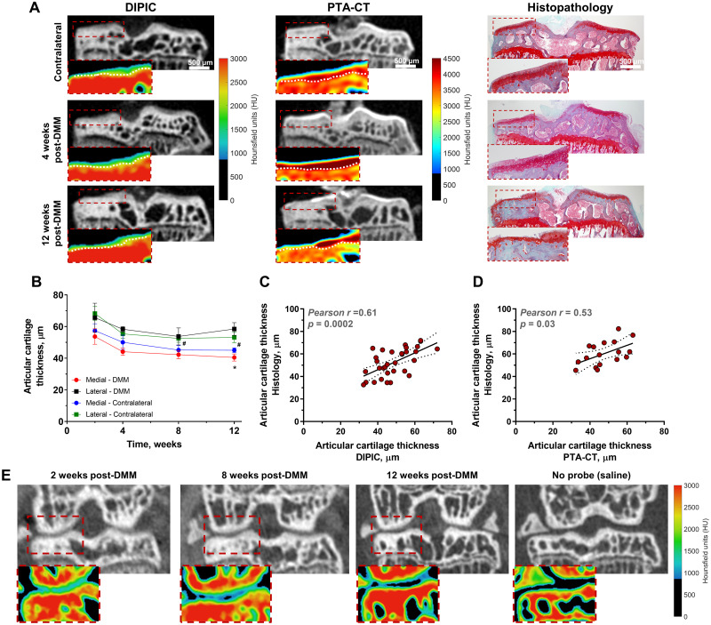
To confer radiopacity to cartilage contrast agents, the naturally occurring tyrosine derivative 3,5-diiodo-L-tyrosine (DIT) was introduced into a selective peptide for type II collagen. Synthetic DIT peptide derivatives were synthesised by Fmoc-based solid-phase peptide synthesis and binding to ex vivo mouse tibial cartilage evaluated by high-resolution micro-CT. Di-Iodotyrosinated Peptide Imaging of Cartilage (DIPIC) was performed ex vivo and in vivo 4, 8 and 12 weeks in mice after induction of OA by destabilisation of the medial meniscus (DMM). Finally, human osteochondral plugs were imaged ex vivo using DIPIC.

RESULTS

Fifteen DIT peptides were synthesised and tested, yielding seven leads with varying cartilage binding strengths. DIPIC visualised ex vivo murine articular cartilage comparably to the ex vivo contrast agent phosphotungstic acid. Intra-articular injection of contrast agent followed by in vivo DIPIC enabled delineation of damaged murine articular cartilage. Finally, the translational potential of the contrast agent was confirmed by visualisation of ex vivo human cartilage explants.

CONCLUSION

DIPIC has reduction and refinement implications in OA animal research and potential clinical translation to imaging human disease.

摘要

背景

现有的磁共振成像(MRI)和新兴的 X 射线对比剂可用于非侵入性成像关节软骨,但它们依赖于与带负电荷的蛋白聚糖的非选择性静电相互作用。由于蛋白聚糖的高周转率,这些对比剂在骨关节炎(OA)等疾病中的预后应用有限。为了克服这一限制,我们开发了一种针对软骨中 II 型胶原大分子的放射性对比剂,并将其用于监测 OA 小鼠模型中的疾病进展。

方法

为了使软骨对比剂具有放射不透性,将天然存在的酪氨酸衍生物 3,5-二碘-L-酪氨酸(DIT)引入到针对 II 型胶原的选择性肽中。通过基于 Fmoc 的固相肽合成合成了 DIT 肽衍生物,并通过高分辨率 micro-CT 评估其在离体小鼠胫骨软骨上的结合情况。在通过内侧半月板不稳定(DMM)诱导 OA 后 4、8 和 12 周,在小鼠体内和体内进行了 Di-Iodotyrosinated Peptide Imaging of Cartilage(DIPIC)。最后,使用 DIPIC 对离体人骨软骨插件进行成像。

结果

合成并测试了 15 种 DIT 肽,得到了 7 种具有不同软骨结合强度的先导肽。DIPIC 可与体外对比剂磷钨酸一样清晰地显示出体外小鼠关节软骨。关节内注射对比剂后,再进行体内 DIPIC 可清晰显示出受损的小鼠关节软骨。最后,通过对离体人软骨外植体的成像,证实了该对比剂的转化潜力。

结论

DIPIC 对 OA 动物研究具有减少和精细化的意义,并有潜在的临床转化为人类疾病成像的应用。

https://cdn.ncbi.nlm.nih.gov/pmc/blobs/4647/9089912/246ca0504a9f/pone.0268223.g004.jpg
https://cdn.ncbi.nlm.nih.gov/pmc/blobs/4647/9089912/8d2e2e0e3a8e/pone.0268223.g001.jpg
https://cdn.ncbi.nlm.nih.gov/pmc/blobs/4647/9089912/9d05931fb6bf/pone.0268223.g002.jpg
https://cdn.ncbi.nlm.nih.gov/pmc/blobs/4647/9089912/6c0bd742b56c/pone.0268223.g003.jpg
https://cdn.ncbi.nlm.nih.gov/pmc/blobs/4647/9089912/246ca0504a9f/pone.0268223.g004.jpg

相似文献

1
Imaging articular cartilage in osteoarthritis using targeted peptide radiocontrast agents.使用靶向肽类放射性对比剂对骨关节炎的关节软骨成像。
PLoS One. 2022 May 10;17(5):e0268223. doi: 10.1371/journal.pone.0268223. eCollection 2022.
2
Synchrotron- and laboratory-based X-ray phase-contrast imaging for imaging mouse articular cartilage in the absence of radiopaque contrast agents.同步辐射和实验室基于 X 射线相衬成像在没有不透射线对比剂的情况下对小鼠关节软骨成像。
Philos Trans A Math Phys Eng Sci. 2014 Jan 27;372(2010):20130127. doi: 10.1098/rsta.2013.0127. Print 2014 Mar 6.
3
Rapid, automated imaging of mouse articular cartilage by microCT for early detection of osteoarthritis and finite element modelling of joint mechanics.通过微型计算机断层扫描对小鼠关节软骨进行快速自动成像,用于骨关节炎的早期检测和关节力学的有限元建模。
Osteoarthritis Cartilage. 2014 Oct;22(10):1419-28. doi: 10.1016/j.joca.2014.07.014.
4
Application of autofluorescence robotic histology for quantitative evaluation of the 3-dimensional morphology of murine articular cartilage.自体荧光机器人组织学在小鼠关节软骨三维形态定量评估中的应用。
Microsc Res Tech. 2017 Dec;80(12):1351-1360. doi: 10.1002/jemt.22948. Epub 2017 Sep 30.
5
Varying development of femoral and tibial subchondral bone tissue and their interaction with articular cartilage during progressing osteoarthritis.在进展性骨关节炎中,股骨和胫骨的软骨下骨组织的不同发育及其与关节软骨的相互作用。
Arch Orthop Trauma Surg. 2020 Dec;140(12):1919-1930. doi: 10.1007/s00402-020-03480-w. Epub 2020 May 30.
6
Exogenous fibroblast growth factor 9 attenuates cartilage degradation and aggravates osteophyte formation in post-traumatic osteoarthritis.外源性成纤维细胞生长因子9可减轻创伤后骨关节炎中的软骨降解并加重骨赘形成。
Osteoarthritis Cartilage. 2016 Dec;24(12):2181-2192. doi: 10.1016/j.joca.2016.07.005. Epub 2016 Jul 27.
7
Tracking Osteoarthritis Progress through Cationic Nanoprobe-Enhanced Photoacoustic Imaging of Cartilage.通过阳离子纳米探针增强光声成象技术对软骨进行骨关节炎的进程追踪。
Acta Biomater. 2020 Jun;109:153-162. doi: 10.1016/j.actbio.2020.04.001. Epub 2020 Apr 24.
8
Anionic Contrast-Enhanced MicroCT Imaging Correlates with Biochemical and Histological Evaluations of Osteoarthritic Articular Cartilage.阴离子对比增强 microCT 成像与骨关节炎关节软骨的生化和组织学评估相关。
Cartilage. 2021 Dec;13(2_suppl):1388S-1397S. doi: 10.1177/1947603520924748. Epub 2020 May 26.
9
Local intra-articular injection of rapamycin delays articular cartilage degeneration in a murine model of osteoarthritis.在骨关节炎小鼠模型中,关节内局部注射雷帕霉素可延缓关节软骨退变。
Arthritis Res Ther. 2014 Nov 17;16(6):482. doi: 10.1186/s13075-014-0482-4.
10
Role of Lipocalin-Type Prostaglandin D Synthase in Experimental Osteoarthritis.脂联素型前列腺素D合成酶在实验性骨关节炎中的作用
Arthritis Rheumatol. 2020 Sep;72(9):1524-1533. doi: 10.1002/art.41297. Epub 2020 Jul 30.

引用本文的文献

1
Modular iodinated carboxybetaine copolymers as charge-sensitive contrast agents for the detection of cartilage degradation.模块化碘化羧基甜菜碱共聚物作为用于检测软骨降解的电荷敏感型造影剂。
Mater Today Bio. 2024 Oct 26;29:101302. doi: 10.1016/j.mtbio.2024.101302. eCollection 2024 Dec.
2
Revealing Detailed Cartilage Function Through Nanoparticle Diffusion Imaging: A Computed Tomography & Finite Element Study.通过纳米粒子扩散成像揭示详细的软骨功能:计算机断层扫描和有限元研究。
Ann Biomed Eng. 2024 Sep;52(9):2584-2595. doi: 10.1007/s10439-024-03552-7. Epub 2024 Jul 16.
3
Biomechanics of the Human Osteochondral Unit: A Systematic Review.

本文引用的文献

1
Propagation phase-contrast micro-computed tomography allows laboratory-based three-dimensional imaging of articular cartilage down to the cellular level.相衬增强的微观 CT 可以对关节软骨进行基于实验室的三维成像,达到细胞水平。
Osteoarthritis Cartilage. 2020 Jan;28(1):102-111. doi: 10.1016/j.joca.2019.10.007. Epub 2019 Oct 31.
2
[ F]-Sodium Fluoride PET/MR Imaging for Bone-Cartilage Interactions in Hip Osteoarthritis: A Feasibility Study.[F]-氟化钠 PET/MR 成像在髋关节骨关节炎骨-软骨相互作用中的可行性研究。
J Orthop Res. 2019 Dec;37(12):2671-2680. doi: 10.1002/jor.24443. Epub 2019 Aug 30.
3
Use of CT Vs. MRI for Diagnosis of Hip or Pelvic Fractures in Elderly Patients After Low Energy Trauma.
人体骨软骨单元的生物力学:系统评价
Materials (Basel). 2024 Apr 8;17(7):1698. doi: 10.3390/ma17071698.
低能量创伤后老年患者髋部或骨盆骨折诊断中CT与MRI的应用比较
Iowa Orthop J. 2019;39(1):179-183.
4
Osteoarthritis development related to cartilage quality-the prognostic value of dGEMRIC after anterior cruciate ligament injury.骨关节炎的发生与软骨质量有关——前交叉韧带损伤后 dGEMRIC 的预后价值。
Osteoarthritis Cartilage. 2019 Nov;27(11):1647-1652. doi: 10.1016/j.joca.2019.06.012. Epub 2019 Jul 4.
5
Mechanoadaptation: articular cartilage through thick and thin.力学适应性:关节软骨的厚薄之道。
J Physiol. 2019 Mar;597(5):1271-1281. doi: 10.1113/JP275451. Epub 2018 Jul 29.
6
Imaging Bone-Cartilage Interactions in Osteoarthritis Using [F]-NaF PET-MRI.使用[F]-氟化钠正电子发射断层扫描-磁共振成像技术对骨关节炎中的骨-软骨相互作用进行成像。
Mol Imaging. 2016 Jan 1;15:1-12. doi: 10.1177/1536012116683597.
7
Automated assessment of bone changes in cross-sectional micro-CT studies of murine experimental osteoarthritis.小鼠实验性骨关节炎横断面显微CT研究中骨变化的自动评估
PLoS One. 2017 Mar 23;12(3):e0174294. doi: 10.1371/journal.pone.0174294. eCollection 2017.
8
Effects of Articular Cartilage Constituents on Phosphotungstic Acid Enhanced Micro-Computed Tomography.关节软骨成分对磷钨酸增强型微型计算机断层扫描的影响。
PLoS One. 2017 Jan 30;12(1):e0171075. doi: 10.1371/journal.pone.0171075. eCollection 2017.
9
In Vivo Contrast-Enhanced Cone Beam CT Provides Quantitative Information on Articular Cartilage and Subchondral Bone.体内对比增强锥形束CT提供有关关节软骨和软骨下骨的定量信息。
Ann Biomed Eng. 2017 Mar;45(3):811-818. doi: 10.1007/s10439-016-1730-3. Epub 2016 Sep 19.
10
Ageing and the pathogenesis of osteoarthritis.衰老与骨关节炎的发病机制
Nat Rev Rheumatol. 2016 Jul;12(7):412-20. doi: 10.1038/nrrheum.2016.65. Epub 2016 May 19.