Savic Dragana, Pedoia Valentina, Seo Youngho, Yang Jaewon, Bucknor Matt, Franc Benjamin L, Majumdar Sharmila
1 Department of Radiology and Biomedical Imaging, University of California, San Francisco, San Francisco, CA, USA.
2 Department of Physiology, Anatomy and Genetics, University of Oxford, Oxford, United Kingdom.
Mol Imaging. 2016 Jan 1;15:1-12. doi: 10.1177/1536012116683597.
Simultaneous positron emission tomography-magnetic resonance imaging (PET-MRI) is an emerging technology providing both anatomical and functional images without increasing the scan time. Compared to the traditional PET/computed tomography imaging, it also exposes the patient to significantly less radiation and provides better anatomical images as MRI provides superior soft tissue characterization. Using PET-MRI, we aim to study interactions between cartilage composition and bone function simultaneously, in knee osteoarthritis (OA).
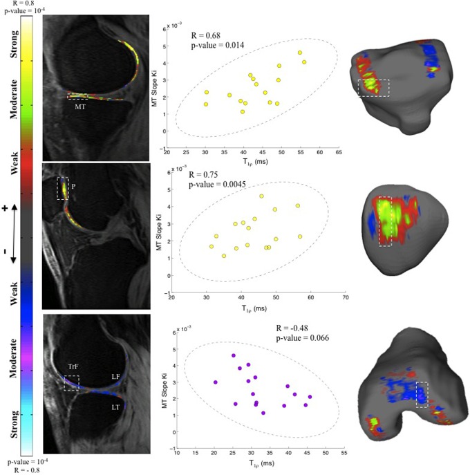
In this article, bone turnover and remodeling was studied using [F]-sodium fluoride (NaF) PET data. Quantitative MR-derived T relaxation times characterized the biochemical cartilage degeneration. Sixteen participants with early signs of OA of the knee received intravenous injections of [F]-NaF at the onset of PET-MR image acquisition. Regions of interest were identified, and kinetic analysis of dynamic PET data provided the rate of uptake ( K) and the normalized uptake (standardized uptake value) of [F]-NaF in the bone. Morphological MR images and quantitative voxel-based T maps of cartilage were obtained using an atlas-based registration technique to segment cartilage automatically. Voxel-by-voxel statistical parameter mapping was used to investigate the relationship between bone and cartilage.
Increases in cartilage T, indicating degenerative changes, were associated with increased turnover in the adjoining bone but reduced turnover in the nonadjoining compartments. Associations between pain and increased bone uptake were seen in the absence of morphological lesions in cartilage, but the relationship was reversed in the presence of incident cartilage lesions.
This study shows significant cartilage and bone interactions in OA of the knee joint using simultaneous [F]-NaF PET-MR, the first in human study. These observations highlight the complex biomechanical and biochemical interactions in the whole knee joint in OA, which potentially could help assess therapeutic targets in treating OA.
同时正电子发射断层扫描 - 磁共振成像(PET - MRI)是一种新兴技术,可在不增加扫描时间的情况下提供解剖学和功能图像。与传统的PET /计算机断层扫描成像相比,它使患者接受的辐射显著减少,并且由于MRI提供了卓越的软组织特征,所以能提供更好的解剖图像。我们旨在使用PET - MRI同时研究膝关节骨关节炎(OA)中软骨成分与骨功能之间的相互作用。
在本文中,使用[F] - 氟化钠(NaF)PET数据研究骨转换和重塑。定量MR衍生的T弛豫时间表征了生化性软骨退变。16名有膝关节OA早期迹象的参与者在PET - MR图像采集开始时接受了[F] - NaF静脉注射。确定感兴趣区域,动态PET数据的动力学分析提供了[F] - NaF在骨中的摄取率(K)和标准化摄取(标准化摄取值)。使用基于图谱的配准技术自动分割软骨,获得形态学MR图像和基于体素的软骨定量T图。逐体素统计参数映射用于研究骨与软骨之间的关系。
软骨T增加表明存在退行性变化,这与相邻骨的转换增加相关,但与不相邻区域的转换减少相关。在软骨无形态学病变的情况下,疼痛与骨摄取增加之间存在关联,但在出现新的软骨病变时这种关系则相反。
本研究首次在人体研究中表明,使用同时进行的[F] - NaF PET - MR可发现膝关节OA中软骨与骨之间存在显著相互作用。这些观察结果突出了OA整个膝关节中复杂的生物力学和生化相互作用,这可能有助于评估OA治疗的靶点。