Chen Jingli, Xue Kangkang, Yang Meng, Wang Kefan, Xu Yinhuan, Wen Baohong, Cheng Jingliang, Han Shaoqiang, Wei Yarui
Department of Magnetic Resonance Imaging, The First Affiliated Hospital of Zhengzhou University, Zhengzhou, China.
Front Neurosci. 2022 Apr 25;16:821078. doi: 10.3389/fnins.2022.821078. eCollection 2022.
Auditory verbal hallucinations (AVHs) are a major symptom of schizophrenia and are connected with impairments in auditory and speech-related networks. In schizophrenia with AVHs, alterations in resting-state cerebral blood flow (CBF) and functional connectivity have been described. However, the neurovascular coupling alterations specific to first-episode drug-naïve schizophrenia (FES) patients with AVHs remain unknown.
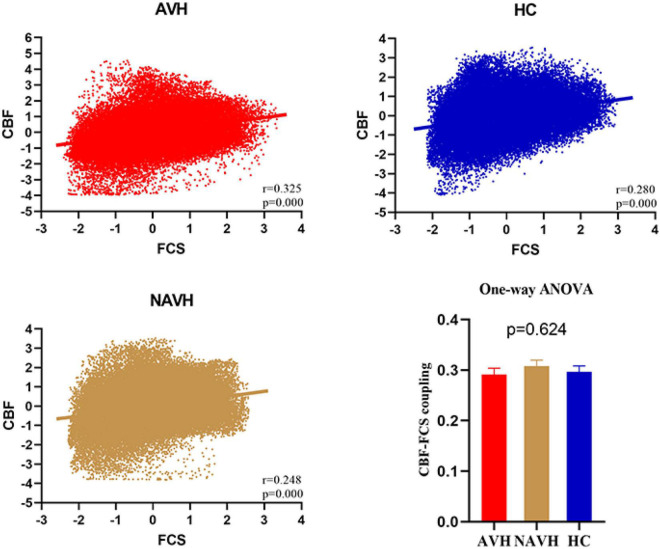
Resting-state functional MRI and arterial spin labeling (ASL) was performed on 46 first-episode drug-naïve schizophrenia (FES) patients with AVHs (AVH), 39 FES drug-naïve schizophrenia patients without AVHs (NAVH), and 48 healthy controls (HC). Then we compared the correlation between the CBF and functional connection strength (FCS) of the entire gray matter between the three groups, as well as the CBF/FCS ratio of each voxel. Correlation analyses were performed on significant results between schizophrenia patients and clinical measures scale.
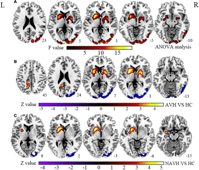
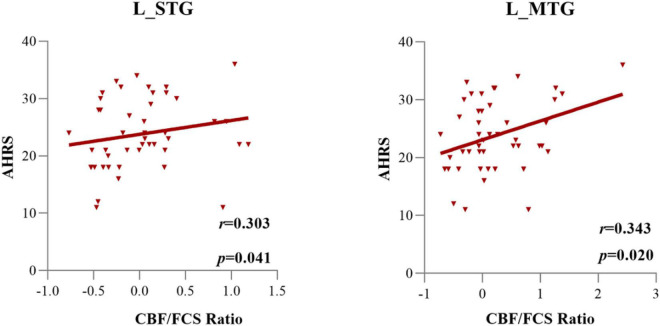
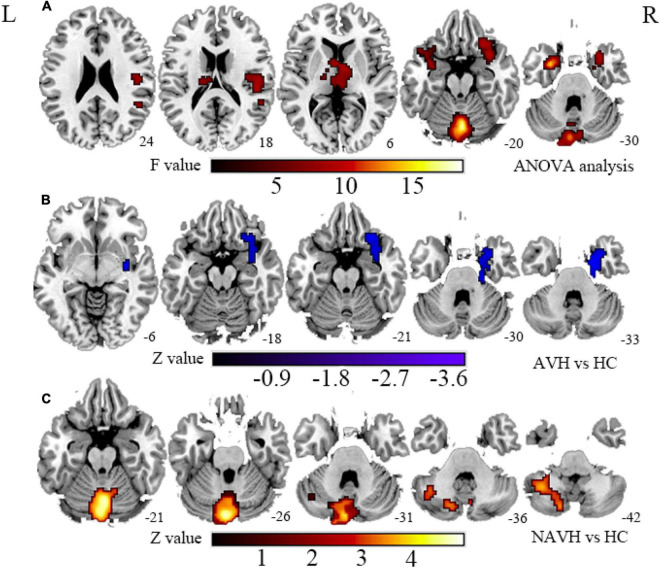
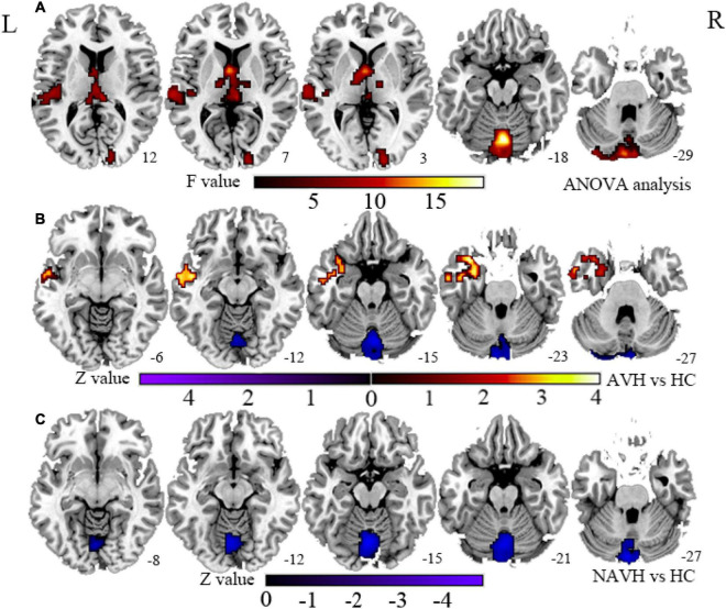
The CBF/FCS ratio was reduced in the cognitive and emotional brain regions in both the AVH and NAVH groups, primarily in the crus I/II, vermis VI/VII, and cerebellum VI. In the AVH group compared with the HC group, the CBF/FCS ratio was higher in auditory perception and language-processing areas, primarily the left superior and middle temporal gyrus (STG/MTG). The CBF/FCS ratio in the left STG and left MTG positively correlates with the score of the Auditory Hallucination Rating Scale in AVH patients.
These findings point to the difference in neurovascular coupling failure between AVH and NAVH patients. The dysfunction of the forward model based on the predictive and computing role of the cerebellum may increase the excitability in the auditory cortex, which may help to understand the neuropathological mechanism of AVHs.
幻听是精神分裂症的主要症状,与听觉及言语相关网络的损伤有关。在伴有幻听的精神分裂症患者中,静息态脑血流量(CBF)和功能连接的改变已有报道。然而,首发未用药精神分裂症(FES)伴幻听患者的神经血管耦合改变仍不清楚。
对46例首发未用药的伴有幻听的精神分裂症(FES)患者(AVH组)、39例首发未用药的无幻听精神分裂症患者(NAVH组)和48名健康对照者(HC组)进行静息态功能磁共振成像和动脉自旋标记(ASL)检查。然后比较三组之间全脑灰质的CBF与功能连接强度(FCS)的相关性,以及每个体素的CBF/FCS比值。对精神分裂症患者与临床量表之间的显著结果进行相关性分析。
AVH组和NAVH组认知和情感脑区的CBF/FCS比值均降低,主要位于小脑脚I/II、蚓部VI/VII和小脑叶VI。与HC组相比,AVH组听觉感知和语言处理区域的CBF/FCS比值更高,主要位于左侧颞上回和颞中回(STG/MTG)。AVH患者左侧STG和左侧MTG的CBF/FCS比值与幻听评定量表评分呈正相关。
这些发现表明AVH和NAVH患者在神经血管耦合功能障碍方面存在差异。基于小脑预测和计算作用的前向模型功能障碍可能会增加听觉皮层的兴奋性,这可能有助于理解幻听的神经病理机制。