Pregler Benedikt, Beyer Lukas Philipp, Platz Batista da Silva Natascha, Steer Sebastian, Zeman Florian, Popp Daniel, Stroszczynski Christian, Müller-Wille René
Department of Radiology, Ernst von Bergmann Klinikum Potsdam, 14467 Potsdam, Germany.
Department of Radiology, University Medical Center Regensburg, 93053 Regensburg, Germany.
J Clin Med. 2022 Apr 29;11(9):2502. doi: 10.3390/jcm11092502.
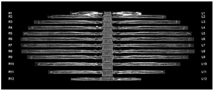
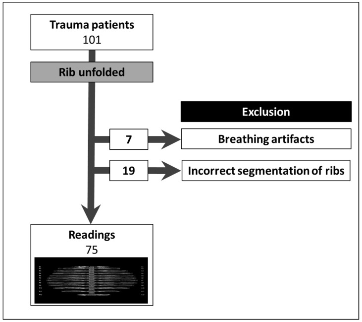
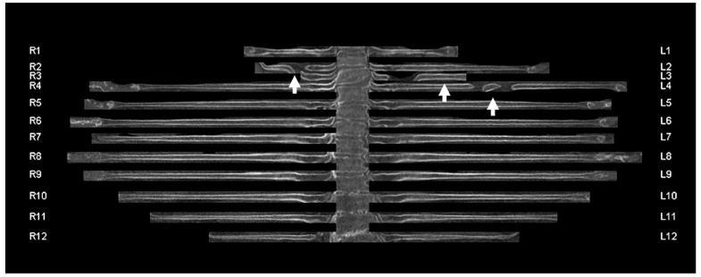
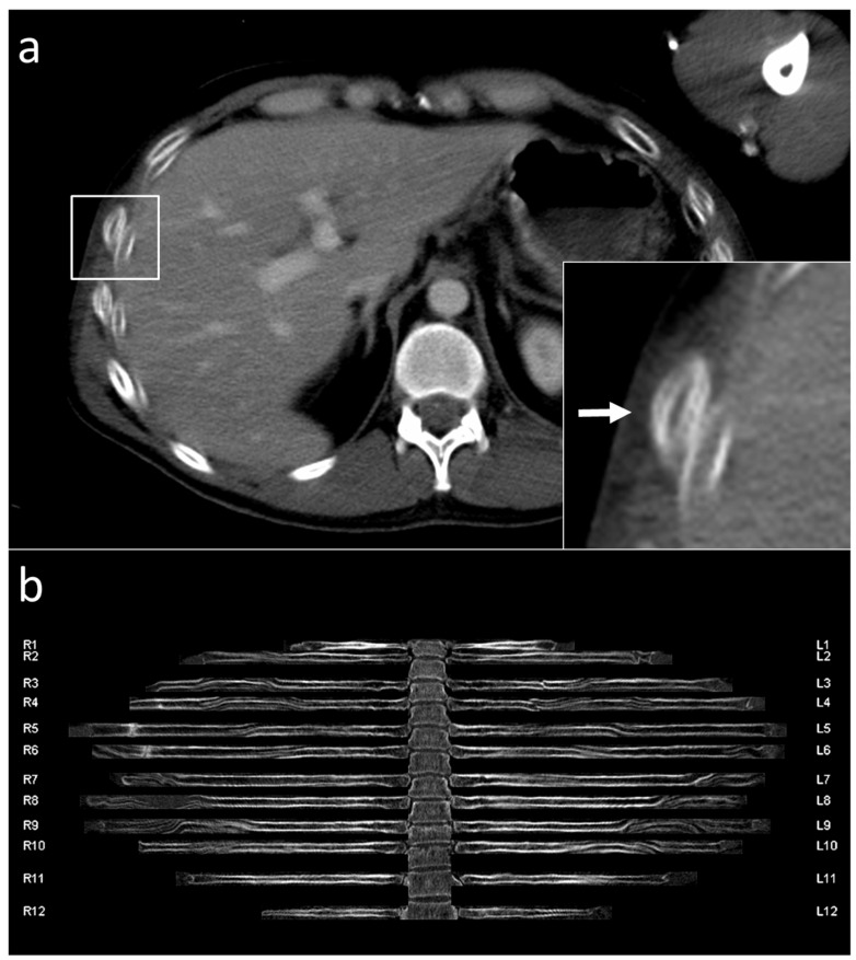
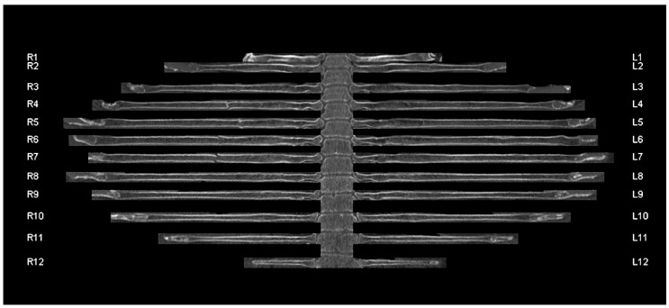
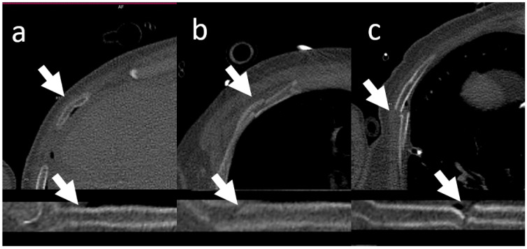
Introduction: The fast and accurate diagnosis of rib fractures in polytrauma patients is important to reduce the mortality rate and relieve long-term pain and complications. Aim: To evaluate the diagnostic accuracy and potential time savings using automatic rib segmentation and a curved, unfolded view for the detection of rib fractures in trauma patients. Methods: The multidetector computed tomography raw data of 101 consecutive polytrauma patients (72 men; mean age 45 years, age range 17 to 84 years) admitted to a university hospital were retrospectively post-processed to generate a curved, unfolded view of the rib cage. No manual corrections were performed. Patients with reconstruction errors and movement artifacts were excluded from further analysis. All fractures were identified and classified by the study coordinator using the original data set. Two readers (reader 1 and reader 2) evaluated the original axial sections and the unfolded view, separately. The fracture locations, fracture type, and reading times were recorded. Sensitivity and specificity were calculated on a per-rib basis using a ratio estimator. Cohen’s Kappa was calculated as an index of inter-rater agreement. Results: 26 of 101 patients (25.7%) were excluded from further analysis owing to breathing artifacts (6.9%) or incorrect centerline computation in the unfolded view (18.8%). In total, 107 (5.9%) of 1800 ribs were fractured in 25 (33%) of 75 patients. The unfolded view had a sensitivity/specificity of 81%/100% (reader 1) and 71%/100% (reader 2) compared to 94%/100% (reader 1; p = 0.002/p = 0.754) and 63%/99% (reader 2; p < 0.001/p = 0.002). The sensitivity (reader 1; reader 2) was poor for buckled fractures (31%; 38%), moderate for undislocated fractures (78%; 62%), and good for dislocated fractures (94%; 90%). The assessment of the unfolded view was performed significantly faster than that of the original layers (19.5 ± 9.4 s vs. 68.6 ± 32.4 s by reader 1 (p < 0.001); 24.1 ± 9.5 s vs. 40.2 ± 12.7 s by reader 2 (p < 0.001)). Both readers demonstrated a very high interobserver agreement for the unfolded view (κ = 0.839) but only a moderate agreement for the original view (κ = 0.529). Conclusion: Apart from a relatively high number of incorrect centerline reconstructions, the unfolded view of the rib cage allows a faster diagnosis of dislocated rib fractures.
快速准确地诊断多发伤患者的肋骨骨折对于降低死亡率、缓解长期疼痛及并发症至关重要。目的:评估使用自动肋骨分割和曲面展开视图检测创伤患者肋骨骨折的诊断准确性及潜在的时间节省情况。方法:对一所大学医院收治的101例连续多发伤患者(72例男性;平均年龄45岁,年龄范围17至84岁)的多排螺旋CT原始数据进行回顾性后处理,以生成胸廓的曲面展开视图。未进行人工校正。将存在重建错误和运动伪影的患者排除在进一步分析之外。研究协调员使用原始数据集识别并分类所有骨折。两名阅片者(阅片者1和阅片者2)分别评估原始轴位图像和展开视图。记录骨折位置、骨折类型及阅片时间。使用比率估计器按每根肋骨计算敏感度和特异度。计算Cohen's Kappa作为阅片者间一致性的指标。结果:101例患者中有26例(25.7%)因呼吸伪影(6.9%)或展开视图中中心线计算错误(18.8%)被排除在进一步分析之外。75例患者中共有1800根肋骨中的107根(5.9%)发生骨折。与阅片者1的94%/100%(p = 0.002/p = 0.754)和阅片者2的63%/99%(p < 0.001/p = 0.002)相比,展开视图的敏感度/特异度为阅片者1的81%/100%和阅片者2的71%/100%。对于皱折骨折,阅片者1和阅片者2的敏感度较差(分别为31%和38%);对于无移位骨折,敏感度中等(分别为78%和62%);对于移位骨折,敏感度良好(分别为94%和90%)。对展开视图的评估明显快于对原始层面的评估(阅片者1:19.5 ± 9.4秒对68.6 ± 32.4秒,p < 0.001;阅片者2:24.1 ± 9.5秒对40.2 ± 12.7秒,p < 0.001)。两名阅片者对展开视图的观察者间一致性非常高(κ = 0.839),但对原始视图的一致性仅为中等(κ = 0.529)。结论:除了相对较高数量的中心线重建错误外,胸廓的展开视图可更快地诊断移位肋骨骨折。