Ernault Auriane C, Verkerk Arie O, Bayer Jason D, Aras Kedar, Montañés-Agudo Pablo, Mohan Rajiv A, Veldkamp Marieke, Rivaud Mathilde R, de Winter Rosan, Kawasaki Makiri, van Amersfoorth Shirley C M, Meulendijks Eva R, Driessen Antoine H G, Efimov Igor R, de Groot Joris R, Coronel Ruben
Department of Clinical, Experimental Cardiology and Medical Biology, Amsterdam UMC, Amsterdam, The Netherlands.
Department of Clinical, Experimental Cardiology and Medical Biology, Amsterdam UMC, Amsterdam, The Netherlands.
Heart Rhythm. 2022 Sep;19(9):1461-1470. doi: 10.1016/j.hrthm.2022.05.011. Epub 2022 May 12.
Epicardial adipose tissue (EAT) accumulation is associated with cardiac arrhythmias. The effect of EAT secretome (EATs) on cardiac electrophysiology remains largely unknown.
The purpose of this study was to investigate the arrhythmogenicity of EATs and its underlying molecular and electrophysiological mechanisms.
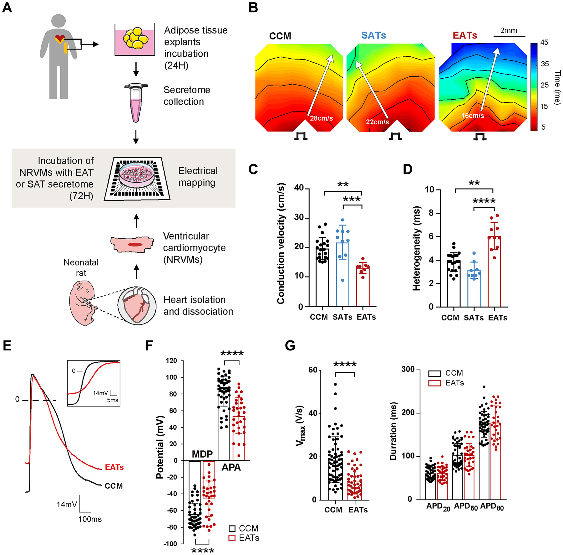
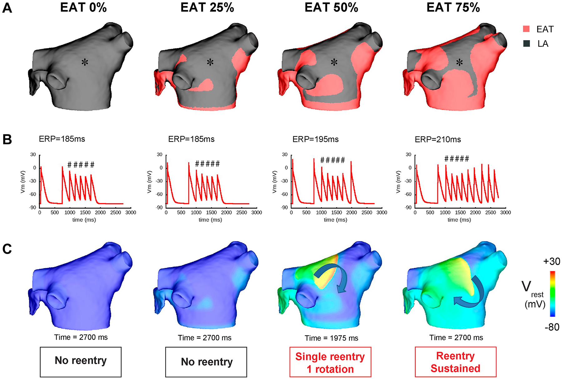
We collected atrial EAT and subcutaneous adipose tissue (SAT) from 30 patients with atrial fibrillation (AF), and EAT from 3 donors without AF. The secretome was collected after a 24-hour incubation of the adipose tissue explants. We cultured neonatal rat ventricular myocytes (NRVMs) with EATs, subcutaneous adipose tissue secretome (SATs), and cardiomyocytes conditioned medium (CCM) for 72 hours. We implemented the electrophysiological changes observed after EATs incubation into a model of human left atrium and tested arrhythmia inducibility.
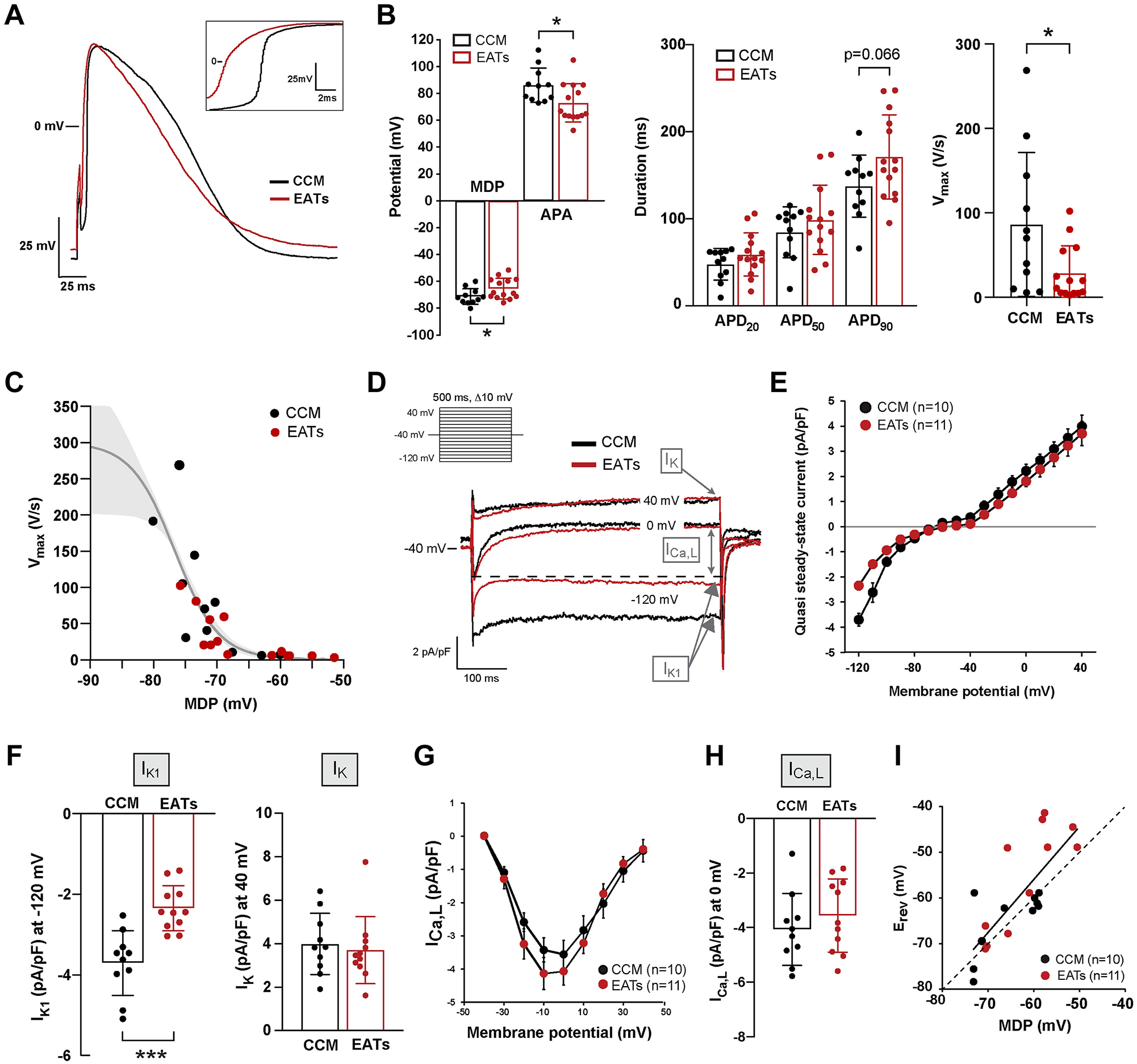
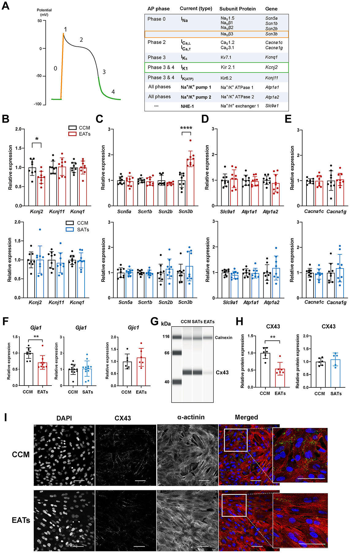
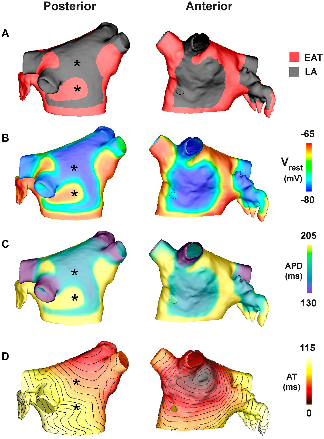
Incubation of NRVMs with EATs decreased expression of the potassium channel subunit Kcnj2 by 26% and correspondingly reduced the inward rectifier K current I by 35% compared to incubation with CCM, resulting in a depolarized resting membrane of cardiomyocytes. EATs decreased expression of connexin43 (29% mRNA, 46% protein) in comparison to CCM. Cells incubated with SATs showed no significant differences in Kcnj2 or Gja1 expression in comparison to CCM, and their resting potential was not depolarized. Cardiomyocytes incubated with EATs showed reduced conduction velocity and increased conduction heterogeneity compared to SATs and CCM. Computer modeling of human left atrium revealed that the electrophysiological changes induced by EATs promote sustained reentrant arrhythmias if EAT partially covers the myocardium.
EAT slows conduction, depolarizes the resting potential, alters electrical cell-cell coupling, and facilitates reentrant arrhythmias.
心外膜脂肪组织(EAT)堆积与心律失常有关。EAT分泌组(EATs)对心脏电生理学的影响在很大程度上仍不清楚。
本研究旨在探讨EATs的致心律失常性及其潜在的分子和电生理机制。
我们收集了30例房颤(AF)患者的心房EAT和皮下脂肪组织(SAT),以及3例无AF供体的EAT。脂肪组织外植体孵育24小时后收集分泌组。我们将新生大鼠心室肌细胞(NRVMs)与EATs、皮下脂肪组织分泌组(SATs)和心肌细胞条件培养基(CCM)培养72小时。我们将EATs孵育后观察到的电生理变化应用于人类左心房模型,并测试心律失常的诱发性。
与CCM孵育相比,用EATs孵育NRVMs使钾通道亚基Kcnj2的表达降低了26%,相应地内向整流钾电流I降低了35%,导致心肌细胞静息膜去极化。与CCM相比,EATs使连接蛋白43的表达降低(mRNA降低29%,蛋白质降低46%)。与CCM相比,用SATs孵育的细胞在Kcnj2或Gja1表达上没有显著差异,其静息电位也没有去极化。与SATs和CCM相比,用EATs孵育的心肌细胞传导速度降低,传导异质性增加。人类左心房的计算机模型显示,如果EAT部分覆盖心肌,EATs诱导的电生理变化会促进持续性折返性心律失常。
EAT减慢传导,使静息电位去极化,改变细胞间电偶联,并促进折返性心律失常。