Translational Imaging in Neurology (ThINK) Basel, Department of Biomedical Engineering, Faculty of Medicine, University Hospital Basel, University of Basel, Basel, Switzerland.
Neurologic Clinic and Policlinic, MS Center and Research Center for Clinical Neuroimmunology and Neuroscience Basel (RC2NB), University Hospital Basel, University of Basel, Basel, Switzerland.
JAMA Neurol. 2022 Jul 1;79(7):682-692. doi: 10.1001/jamaneurol.2022.1025.
The mechanisms driving neurodegeneration and brain atrophy in relapsing multiple sclerosis (RMS) are not completely understood.
To determine whether disability progression independent of relapse activity (PIRA) in patients with RMS is associated with accelerated brain tissue loss.
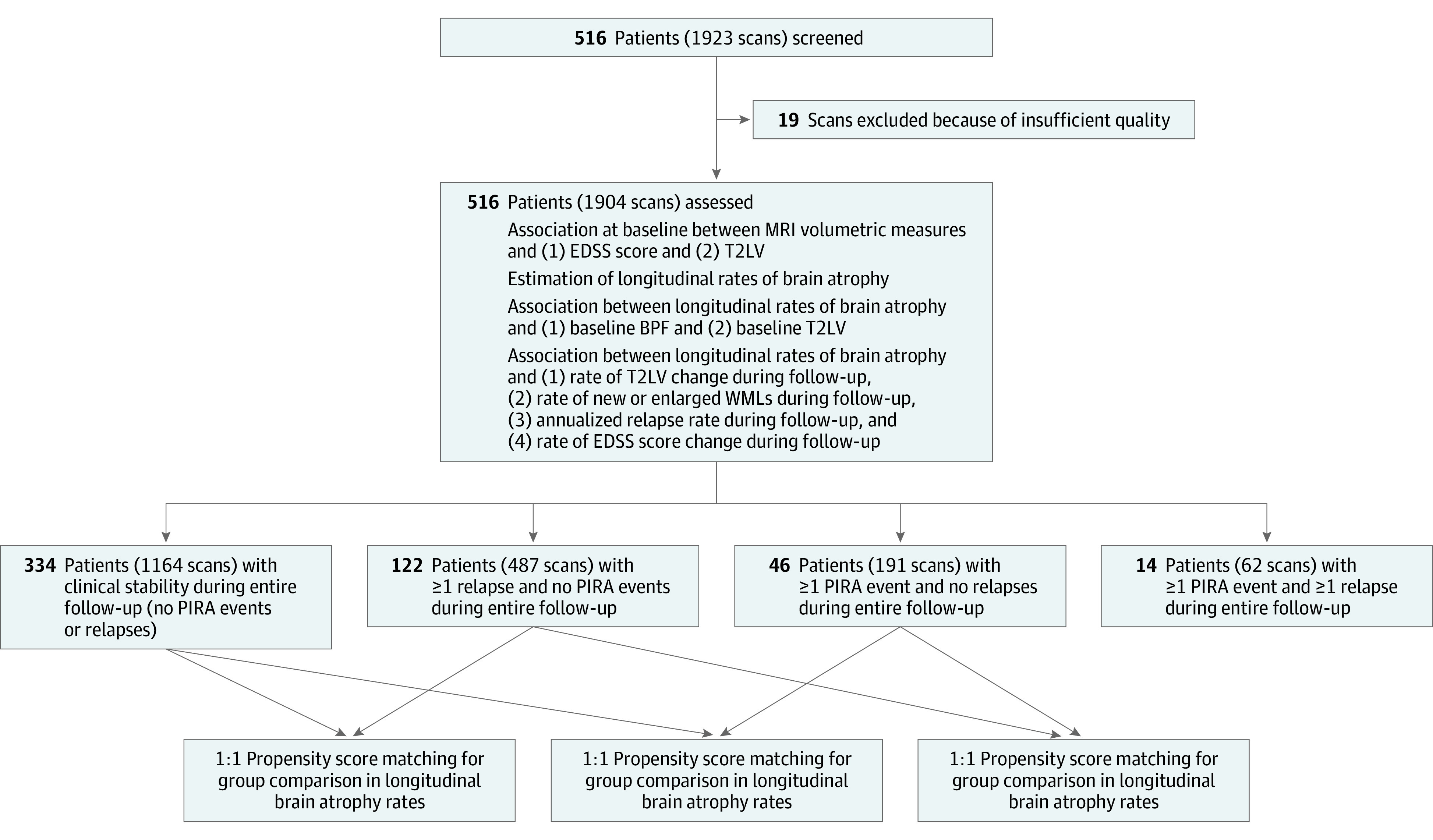
DESIGN, SETTING, AND PARTICIPANTS: In this observational, longitudinal cohort study with median (IQR) follow-up of 3.2 years (2.0-4.9), data were acquired from January 2012 to September 2019 in a consortium of tertiary university and nonuniversity referral hospitals. Patients were included if they had regular clinical follow-up and at least 2 brain magnetic resonance imaging (MRI) scans suitable for volumetric analysis. Data were analyzed between January 2020 and March 2021.
According to the clinical evolution during the entire observation, patients were classified as those presenting (1) relapse activity only, (2) PIRA episodes only, (3) mixed activity, or (4) clinical stability.
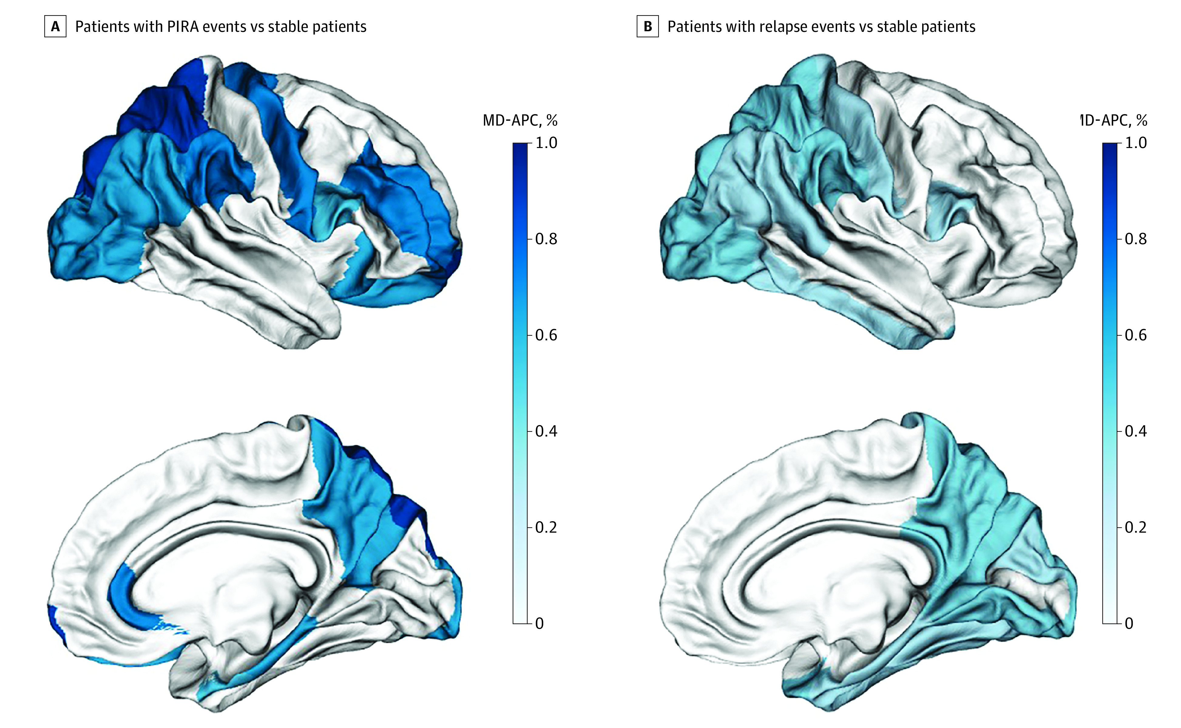
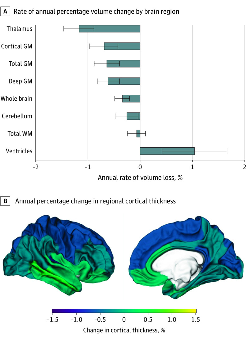
Mean difference in annual percentage change (MD-APC) in brain volume/cortical thickness between groups, calculated after propensity score matching. Brain atrophy rates, and their association with the variables of interest, were explored with linear mixed-effect models.
Included were 1904 brain MRI scans from 516 patients with RMS (67.4% female; mean [SD] age, 41.4 [11.1] years; median [IQR] Expanded Disability Status Scale score, 2.0 [1.5-3.0]). Scans with insufficient quality were excluded (n = 19). Radiological inflammatory activity was associated with increased atrophy rates in several brain compartments, while an increased annualized relapse rate was linked to accelerated deep gray matter (GM) volume loss. When compared with clinically stable patients, patients with PIRA had an increased rate of brain volume loss (MD-APC, -0.36; 95% CI, -0.60 to -0.12; P = .02), mainly driven by GM loss in the cerebral cortex. Patients who were relapsing presented increased whole brain atrophy (MD-APC, -0.18; 95% CI, -0.34 to -0.02; P = .04) with respect to clinically stable patients, with accelerated GM loss in both cerebral cortex and deep GM. No differences in brain atrophy rates were measured between patients with PIRA and those presenting relapse activity.
Our study shows that patients with RMS and PIRA exhibit accelerated brain atrophy, especially in the cerebral cortex. These results point to the need to recognize the insidious manifestations of PIRA in clinical practice and to further evaluate treatment strategies for patients with PIRA in clinical trials.
复发型多发性硬化症(RMS)中导致神经退行性变和脑萎缩的机制尚不完全清楚。
确定 RMS 患者中与复发活动无关的残疾进展(PIRA)是否与加速脑组织损失有关。
设计、设置和参与者:在这项观察性、纵向队列研究中,中位(IQR)随访时间为 3.2 年(2.0-4.9),数据来自 2012 年 1 月至 2019 年 9 月的大学和非大学转诊医院联盟。如果患者有定期的临床随访和至少 2 次适合体积分析的脑磁共振成像(MRI)扫描,则将其纳入研究。数据分析于 2020 年 1 月至 2021 年 3 月进行。
根据整个观察期间的临床演变,患者分为以下几类:(1)仅出现复发活动,(2)仅出现 PIRA 发作,(3)混合活动,或(4)临床稳定。
在进行倾向评分匹配后,计算各组间脑容量/皮质厚度的年百分比变化均值差异(MD-APC)。使用线性混合效应模型探索脑萎缩率及其与感兴趣变量的关系。
纳入了 516 例 RMS 患者的 1904 次脑 MRI 扫描(67.4%为女性;平均[SD]年龄为 41.4[11.1]岁;扩展残疾状态量表评分中位数[IQR]为 2.0[1.5-3.0])。排除了质量不足的扫描(n=19)。放射学炎症活动与多个脑区的萎缩率增加有关,而年化复发率的增加与深部灰质(GM)体积的加速损失有关。与临床稳定的患者相比,PIRA 患者的脑容量损失率增加(MD-APC,-0.36;95%CI,-0.60 至 -0.12;P=0.02),主要是由大脑皮层 GM 损失驱动的。复发患者的全脑萎缩速度加快(MD-APC,-0.18;95%CI,-0.34 至 -0.02;P=0.04),与临床稳定患者相比,大脑皮层和深部 GM 的 GM 损失均加速。在 PIRA 患者和出现复发活动的患者之间,未测量到脑萎缩率的差异。
本研究表明,PIRA 患者的 RMS 表现出加速的脑萎缩,尤其是大脑皮层。这些结果表明,需要在临床实践中认识到 PIRA 的隐匿表现,并在临床试验中进一步评估 PIRA 患者的治疗策略。