Department of Civil, Construction and Environmental Engineering, Center for Engineered Cancer Testbeds, Materials and Nanotechnology Program, North Dakota State University, Fargo, ND, 58108, USA.
Laser Biomedical Research Center, G. R. Harrison Spectroscopy Laboratory, Massachusetts Institute of Technology, MB, 02139, Cambridge, USA.
Sci Rep. 2022 May 16;12(1):8050. doi: 10.1038/s41598-022-11800-w.
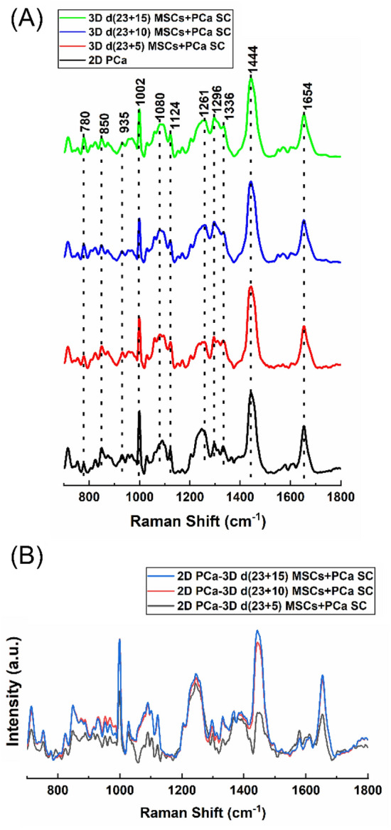
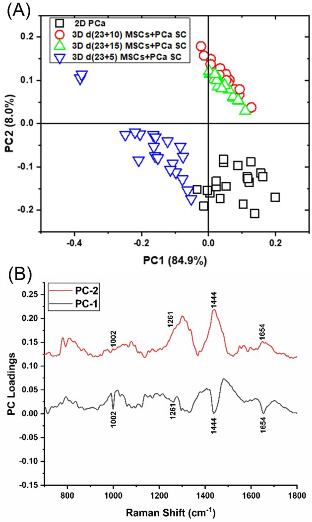
Metastatic prostate cancer colonizes the bone to pave the way for bone metastasis, leading to skeletal complications associated with poor prognosis and morbidity. This study demonstrates the feasibility of Raman imaging to differentiate between cancer cells at different stages of tumorigenesis using a nanoclay-based three-dimensional (3D) bone mimetic in vitro model that mimics prostate cancer bone metastasis. A comprehensive study comparing the classification of as received prostate cancer cells in a two-dimensional (2D) model and cancer cells in a 3D bone mimetic environment was performed over various time intervals using principal component analysis (PCA). Our results showed distinctive spectral differences in Raman imaging between prostate cancer cells and the cells cultured in 3D bone mimetic scaffolds, particularly at 1002, 1261, 1444, and 1654 cm, which primarily contain proteins and lipids signals. Raman maps capture sub-cellular responses with the progression of tumor cells into metastasis. Raman feature extraction via cluster analysis allows for the identification of specific cellular constituents in the images. For the first time, this work demonstrates a promising potential of Raman imaging, PCA, and cluster analysis to discriminate between cancer cells at different stages of metastatic tumorigenesis.
转移性前列腺癌在骨骼中定植,为骨转移铺平道路,导致预后不良和发病率高的骨骼并发症。本研究展示了使用基于纳米粘土的三维(3D)骨仿生体外模型对不同肿瘤发生阶段的癌细胞进行区分的拉曼成像的可行性,该模型模拟了前列腺癌骨转移。使用主成分分析(PCA)在不同时间间隔内对二维(2D)模型中的接收前列腺癌细胞和 3D 骨仿生环境中的癌细胞进行了全面的分类比较研究。我们的结果表明,拉曼成像在前列腺癌细胞和在 3D 骨仿生支架中培养的细胞之间存在明显的光谱差异,特别是在 1002、1261、1444 和 1654 cm 处,这些差异主要包含蛋白质和脂质信号。拉曼图谱捕捉到肿瘤细胞向转移进展时的亚细胞反应。通过聚类分析进行拉曼特征提取可识别图像中的特定细胞成分。这项工作首次证明了拉曼成像、PCA 和聚类分析在区分转移性肿瘤发生不同阶段的癌细胞方面具有很大的潜力。