Department II of Internal Medicine and Center for Molecular Medicine Cologne (CMMC), University of Cologne, Cologne, Germany.
Cologne Excellence Cluster on Cellular Stress Responses in Aging-Associated Diseases (CECAD), University of Cologne, Cologne, Germany.
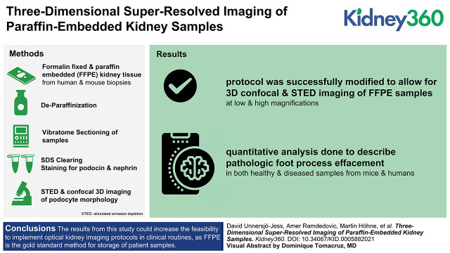
Kidney360. 2021 Dec 1;3(3):446-454. doi: 10.34067/KID.0005882021. eCollection 2022 Mar 31.
Diseases of the glomeruli, the renal filtration units, are a leading cause of progressive kidney disease. Assessment of the ultrastructure of podocytes at the glomerular filtration barrier is essential for diagnosing diverse disease entities, providing insight into the disease pathogenesis, and monitoring treatment responses.
Here we apply previously published sample preparation methods together with stimulated emission depletion and confocal microscopy for resolving nanoscale podocyte substructure. The protocols are modified and optimized in order to be applied to formalin-fixed paraffin-embedded (FFPE) samples.
We successfully modified our protocols to allow for deep three-dimensional stimulated emission depletion and confocal imaging of FFPE kidney tissue with similar staining and image quality compared with our previous approaches. We further show that quantitative analysis can be applied to extract morphometrics from healthy and diseased samples from both mice and humans.
The results from this study could increase the feasibility of implementing optical kidney imaging protocols in clinical routines because FFPE is the gold-standard method for storage of patient samples.
肾小球疾病是导致进行性肾病的主要原因,肾小球是肾脏的过滤单位。评估肾小球滤过屏障的足细胞超微结构对于诊断各种疾病实体、深入了解疾病发病机制以及监测治疗反应至关重要。
在这里,我们应用先前发表的样本制备方法,结合受激发射损耗和共聚焦显微镜,解析纳米级足细胞亚结构。为了将这些方案应用于福尔马林固定石蜡包埋(FFPE)样本,我们对其进行了修改和优化。
我们成功地修改了我们的方案,使 FFPE 肾脏组织能够进行深度三维受激发射损耗和共聚焦成像,与我们之前的方法相比,具有相似的染色和图像质量。我们进一步表明,定量分析可用于从来自小鼠和人类的健康和患病样本中提取形态计量学数据。
这项研究的结果可能会增加在临床常规中实施光学肾脏成像方案的可行性,因为 FFPE 是储存患者样本的金标准方法。