Ustyniak Sergiusz, Stefańczyk Ludomir, Kurnatowska Ilona, Kaczmarska Magdalena, Goździk Maciej
Department of Radiology and Diagnostic Imaging, Medical University of Łódź, Poland.
Department of Internal Medicine and Transplantation Nephrology, 1st Chair of Internal Medicine, Medical University of Łódź, Poland.
Pol J Radiol. 2022 Apr 15;87:e226-e231. doi: 10.5114/pjr.2022.115804. eCollection 2022.
The objectives of our study were to evaluate the changes in the cross-section area of carotid and femoral arteries caused by fluid loss during haemodialysis (HD) and to determine the direction and amount of these changes.
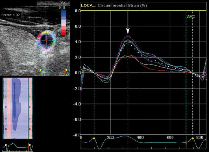
Seventy-four HD patients (28 women and 46 men) were studied. We performed ultrasound exams of the distal common carotid and proximal femoral arteries in each patient before and after a HD session. Recorded exams were analysed using EchoPac software. Arterial cross-section area values were acquired for further analysis.
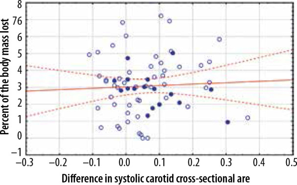
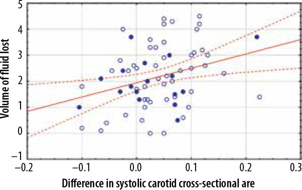
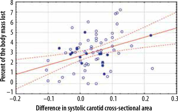
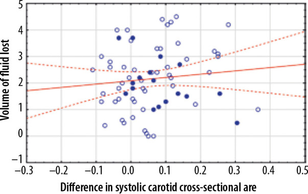
We found a statistically significant decrease in arterial systolic cross-section area values after HD sessions (carotid arteries area before HD equalled 0.6731 cm and 0.6333 cm, = 0.00001 after HD, femoral arteries area before HD equalled 0.8263 cm and 0.7635 cm, = 0.00001 after HD). The decrease of systolic carotid cross-section area correlated with the amount of fluid lost during HD sessions (correlation coefficient of 0.3122, = 0.010) and the percentage of the body mass lost during HD (correlation coefficient of 0.3577, = 0.003). No statistically significant changes were found in the femoral cross-section area.
Our findings suggest that the arterial cross-section area may be used in the assessment of response to body fluid loss. We were able to measure changes due to fluid loss during the HD session. The carotid cross-section values decreased after the procedure and correlated with the amount of fluid lost during the HD session.
我们研究的目的是评估血液透析(HD)期间液体丢失引起的颈动脉和股动脉横截面积的变化,并确定这些变化的方向和量。
对74例HD患者(28例女性和46例男性)进行了研究。我们在每位患者进行HD治疗前后对其颈总动脉远端和股动脉近端进行了超声检查。使用EchoPac软件对记录的检查结果进行分析。获取动脉横截面积值以进行进一步分析。
我们发现HD治疗后动脉收缩期横截面积值有统计学意义的下降(HD前颈动脉面积为0.6731平方厘米,HD后为0.6333平方厘米,P = 0.00001;HD前股动脉面积为0.8263平方厘米,HD后为0.7635平方厘米,P = 0.00001)。颈动脉收缩期横截面积的下降与HD期间丢失的液体量相关(相关系数为0.3122,P = 0.010),也与HD期间体重减轻的百分比相关(相关系数为0.3577,P = 0.003)。股动脉横截面积未发现有统计学意义的变化。
我们的研究结果表明,动脉横截面积可用于评估对体液丢失的反应。我们能够测量HD治疗期间因液体丢失引起的变化。治疗后颈动脉横截面积值下降,并与HD期间丢失的液体量相关。