Department of Surgery, Dijklander Hospital, Hoorn, The Netherlands.
Department of Surgery, UMC, Location AMC, Amsterdam Gastroenterology & Metabolism, University of Amsterdam, Amsterdam, The Netherlands.
Int J Colorectal Dis. 2022 Jun;37(6):1385-1391. doi: 10.1007/s00384-022-04173-z. Epub 2022 May 18.
Radiologic imaging can accurately diagnose acute appendicitis, but little is known about its discriminatory capacity between complicated and uncomplicated appendicitis.
This study aims to investigate the accuracy of imaging in discriminating complicated from uncomplicated appendicitis.
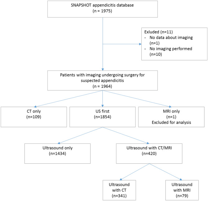
Data was used from the prospective, nationwide, observational SNAPSHOT appendicitis database, including patients with suspected acute appendicitis who were planned for an appendectomy. Usage of ultrasound (US), CT, MRI or a combination was recorded. Radiological reports were used to group for complicated or uncomplicated appendicitis. The reference standard was based on operative and pathological findings. Primary outcomes were sensitivity and specificity in discriminating complicated from uncomplicated appendicitis. Secondary outcomes were diagnostic accuracy results per imaging modality and for the subgroups age, BMI, and sex.
Preoperative imaging was performed in 1964 patients. In 1434 patients (73%), only US was used; in 109 (6%) patients, only CT was used; and 421 (21%) patients underwent US followed by CT or MRI. Overall, imaging workup as practiced, following the national guideline, had a poor sensitivity for complicated appendicitis of only 35%, although specificity was as high as 93%. For US, accuracy for complicated appendicitis was higher in children than in adults; sensitivity 41.2% vs. 26.4% and specificity 94.6% vs. 93.4%, respectively, p = 0.003. For relevant subgroups such as age, sex and BMI, no other differences in the discriminatory performance were found.
A diagnostic workup with stepwise imaging, using a conditional CT or MRI strategy, poorly discriminates between complicated and uncomplicated appendicitis in daily practice.
放射影像学可以准确诊断急性阑尾炎,但对于其在区分复杂型与单纯型阑尾炎方面的鉴别能力知之甚少。
本研究旨在探讨影像学在鉴别复杂型与单纯型阑尾炎方面的准确性。
本研究使用了前瞻性、全国性、观察性的 SNAPSHOT 阑尾炎数据库的数据,纳入计划行阑尾切除术的疑似急性阑尾炎患者。记录了超声(US)、CT、MRI 或联合使用的情况。放射学报告用于将阑尾炎分为复杂型或单纯型。参考标准基于手术和病理发现。主要结局是区分复杂型与单纯型阑尾炎的敏感性和特异性。次要结局是每种影像学方法的诊断准确性结果以及年龄、BMI 和性别亚组的结果。
在 1964 名患者中进行了术前影像学检查。在 1434 名患者(73%)中仅使用 US;在 109 名患者(6%)中仅使用 CT;421 名患者(21%)行 US 后行 CT 或 MRI。总体而言,根据国家指南进行的影像学检查对复杂型阑尾炎的敏感性仅为 35%,特异性高达 93%,其结果较差。对于 US,儿童的复杂型阑尾炎诊断准确性高于成人;敏感性分别为 41.2%和 26.4%,特异性分别为 94.6%和 93.4%,p=0.003。对于年龄、性别和 BMI 等相关亚组,未发现鉴别性能的其他差异。
在日常实践中,使用条件性 CT 或 MRI 策略的逐步影像学检查对区分复杂型与单纯型阑尾炎的能力较差。