Department of Radiology, Kobe University Graduate School of Medicine.
Magn Reson Med Sci. 2023 Oct 1;22(4):435-445. doi: 10.2463/mrms.rev.2021-0152. Epub 2022 May 18.
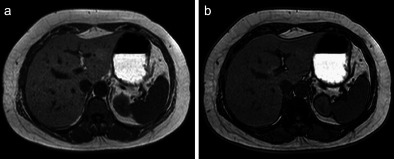
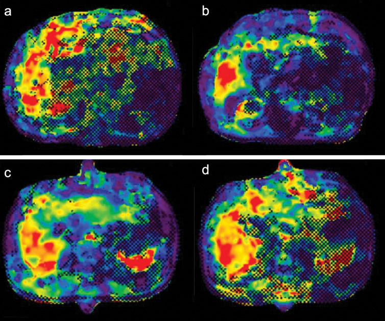
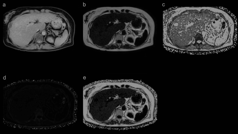
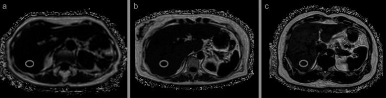
Viral hepatitis was previously the most common cause of chronic liver disease. However, in recent years, nonalcoholic fatty liver disease (NAFLD) cases have been increasing, especially in developed countries. NAFLD is histologically characterized by fat, fibrosis, and inflammation in the liver, eventually leading to cirrhosis and hepatocellular carcinoma. Although biopsy is the gold standard for the assessment of the liver parenchyma, quantitative evaluation methods, such as ultrasound, CT, and MRI, have been reported to have good diagnostic performances. The quantification of liver fat, fibrosis, and inflammation is expected to be clinically useful in terms of the prognosis, early intervention, and treatment response for the management of NAFLD. The aim of this review was to discuss the basics and prospects of MRI-based tissue quantifications of the liver, mainly focusing on proton density fat fraction for the quantification of fat deposition, MR elastography for the quantification of fibrosis, and multifrequency MR elastography for the evaluation of inflammation.
病毒性肝炎以前是慢性肝病的最常见病因。然而,近年来,非酒精性脂肪性肝病(NAFLD)的病例一直在增加,尤其是在发达国家。NAFLD 的组织学特征是肝脏脂肪、纤维化和炎症,最终导致肝硬化和肝细胞癌。虽然肝活检是评估肝实质的金标准,但已有报道称,超声、CT 和 MRI 等定量评估方法具有良好的诊断性能。肝脂肪、纤维化和炎症的定量有望在 NAFLD 的预后、早期干预和治疗反应的临床管理方面具有临床意义。本综述的目的是讨论基于 MRI 的肝脏组织定量的基础和前景,主要集中在质子密度脂肪分数用于脂肪沉积的定量、磁共振弹性成像用于纤维化的定量以及多频磁共振弹性成像用于炎症的评估。