Santiana Leni, Mahmudah Raisa
Department of Radiology, Faculty of Medicine Universitas Padjadjaran, Hasan Sadikin General Hospital, Jl. Pasteur No. 38, Pasteur, Kec. Sukajadi, Bandung, Indonesia.
Radiol Case Rep. 2022 May 9;17(7):2464-2469. doi: 10.1016/j.radcr.2022.04.015. eCollection 2022 Jul.
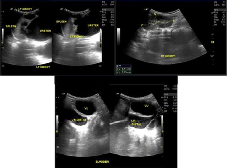
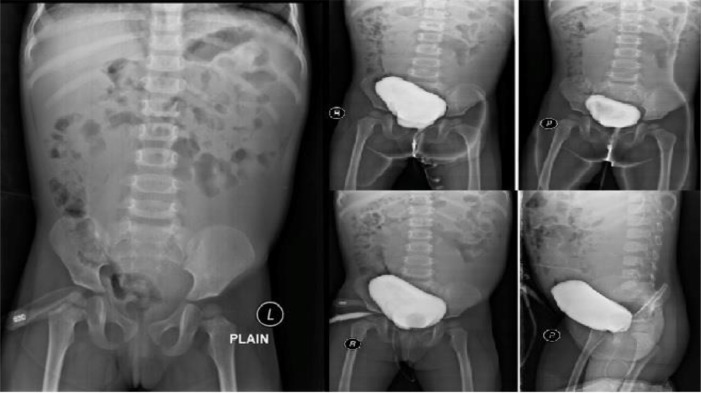
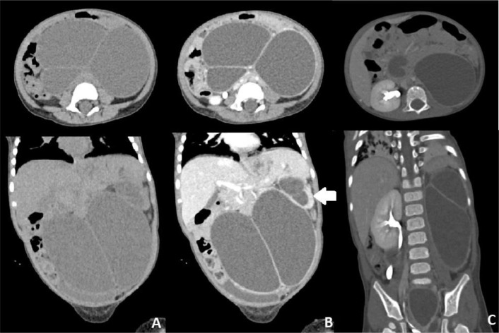
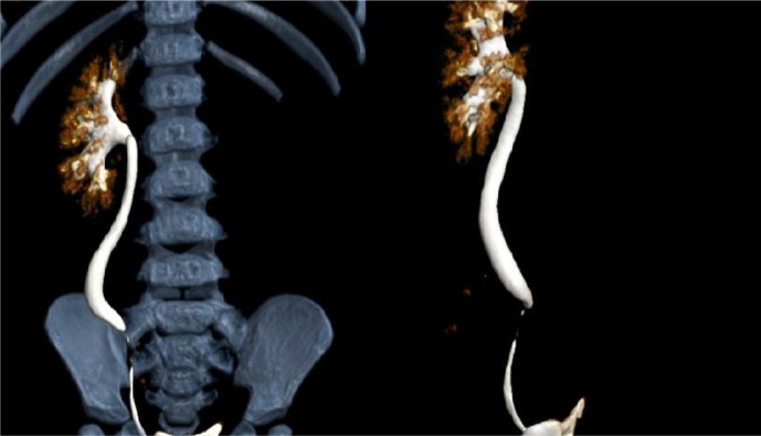
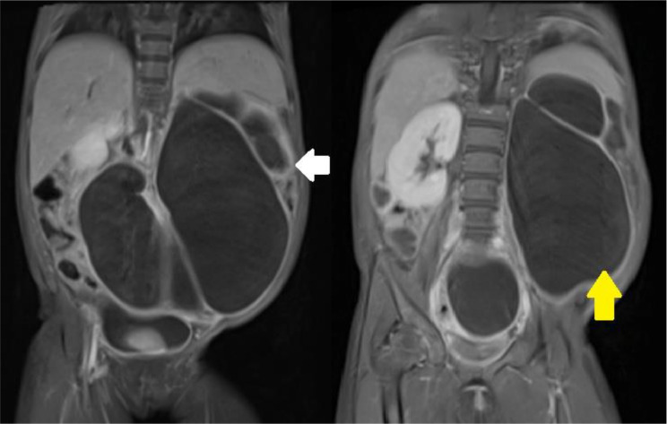
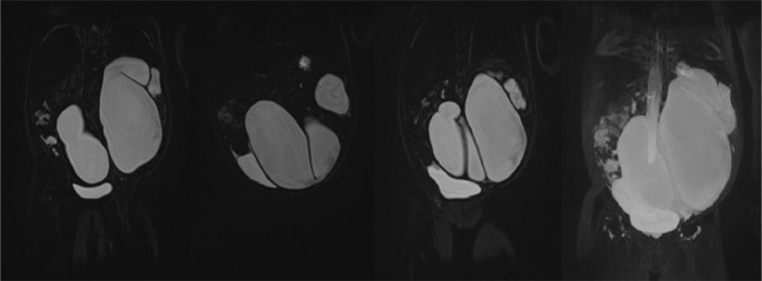
We describe a case of a 2-year-old girl with congenital megaureter presenting as intraabdominal cystic masses. The patient presented with a lump in abdomen that has been getting bigger since birth accompanied by pain. Ultrasonography that was taken when the patient was 2 years old showed a cystic mass with thick septation and pelvocaliectasis of the left kidney. One month after US, patient underwent 3D CT Scan which showed cystic masses in the upper to lower abdomen with no visualization of the normal structure of the left kidney and ureter. Non-contrast MRU that was taken 3 month after the CT Scan showed a thick-walled cystic mass resembling a tortuous tubular mass associated with the pelvocalyceal system without any distal obstruction. VCUG examination that was taken 2 weeks after the non-contrast MRU showed no reflux. This case reports can help clinicians to confirm persistent urinary tract dilatation, exclude the presence of VUR and differentiate primary megaureters from other causes of hydronephrosis including obstruction of the VUJ, posterior urethral valves, and ureterocele from radiological studies.
我们描述了一例2岁先天性巨输尿管女童,表现为腹腔内囊性肿块。该患者自出生以来腹部出现肿块,且不断增大,并伴有疼痛。患者2岁时进行的超声检查显示有一个伴有厚分隔的囊性肿块以及左肾肾盂积水。超声检查后1个月,患者接受了三维CT扫描,结果显示上腹部至下腹部有囊性肿块,未见左肾和输尿管的正常结构。CT扫描后3个月进行的非增强磁共振尿路造影显示一个厚壁囊性肿块,类似与肾盂肾盏系统相关联的迂曲管状肿块,无任何远端梗阻。非增强磁共振尿路造影后2周进行的排尿性膀胱尿道造影检查显示无反流。本病例报告有助于临床医生确认持续性尿路扩张,排除膀胱输尿管反流的存在,并通过影像学研究将原发性巨输尿管与其他肾积水原因(包括输尿管膀胱连接部梗阻、后尿道瓣膜和输尿管囊肿)相鉴别。