Planche Vincent, Manjon José V, Mansencal Boris, Lanuza Enrique, Tourdias Thomas, Catheline Gwenaëlle, Coupé Pierrick
Univ. Bordeaux, CNRS, UMR 5293, Institut des Maladies Neurodégénératives, F-33000 Bordeaux, France.
Instituto de Aplicaciones de las Tecnologías de la Información y de las Comunicaciones Avanzadas (ITACA), Universitat Politècnica de València, Camino de Vera s/n, 46022 Valencia, Spain.
Brain Commun. 2022 Apr 28;4(3):fcac109. doi: 10.1093/braincomms/fcac109. eCollection 2022.
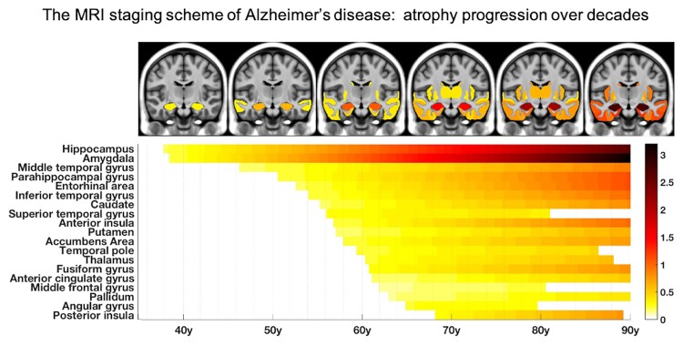
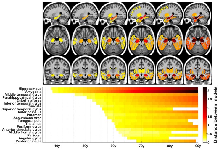
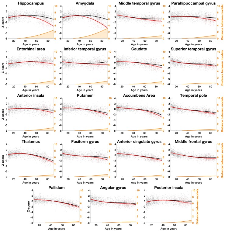
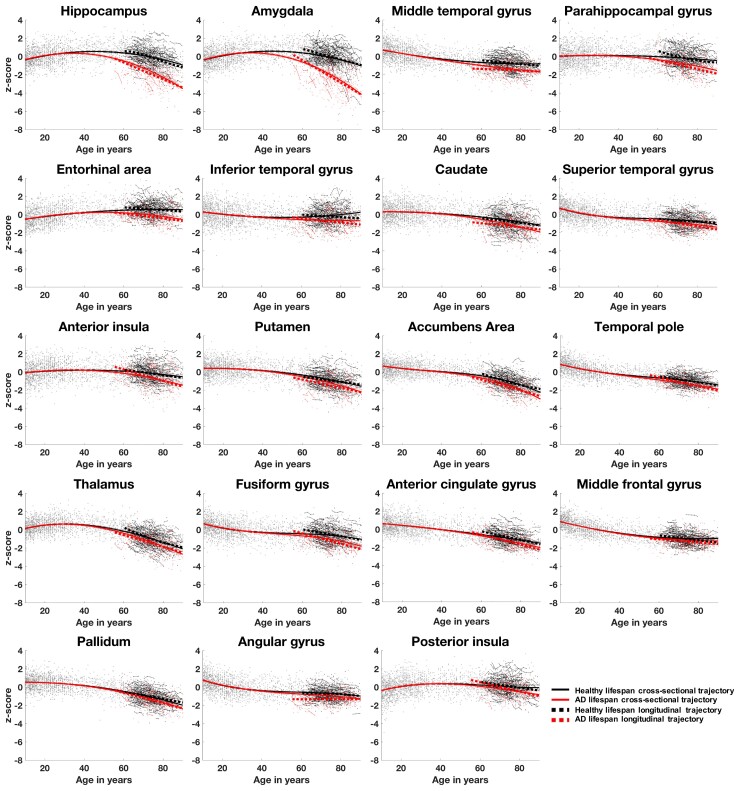
The chronological progression of brain atrophy over decades, from pre-symptomatic to dementia stages, has never been formally depicted in Alzheimer's disease. This is mainly due to the lack of cohorts with long enough MRI follow-ups in cognitively unimpaired young participants at baseline. To describe a spatiotemporal atrophy staging of Alzheimer's disease at the whole-brain level, we built extrapolated lifetime volumetric models of healthy and Alzheimer's disease brain structures by combining multiple large-scale databases ( = 3512 quality controlled MRI from 9 cohorts of subjects covering the entire lifespan, including 415 MRI from ADNI1, ADNI2 and AIBL for Alzheimer's disease patients). Then, we validated dynamic models based on cross-sectional data using external longitudinal data. Finally, we assessed the sequential divergence between normal aging and Alzheimer's disease volumetric trajectories and described the following staging of brain atrophy progression in Alzheimer's disease: (i) hippocampus and amygdala; (ii) middle temporal gyrus; (iii) entorhinal cortex, parahippocampal cortex and other temporal areas; (iv) striatum and thalamus and (v) middle frontal, cingular, parietal, insular cortices and pallidum. We concluded that this MRI scheme of atrophy progression in Alzheimer's disease was close but did not entirely overlap with Braak staging of tauopathy, with a 'reverse chronology' between limbic and entorhinal stages. Alzheimer's disease structural progression may be associated with local tau accumulation but may also be related to axonal degeneration in remote sites and other limbic-predominant associated proteinopathies.
在阿尔茨海默病中,从症状前到痴呆阶段,大脑萎缩在数十年间的时间进程从未被正式描述过。这主要是由于缺乏在基线时认知未受损的年轻参与者中进行足够长时间MRI随访的队列。为了描述阿尔茨海默病在全脑水平的时空萎缩分期,我们通过整合多个大规模数据库(来自9个涵盖整个寿命期的队列的3512份质量控制的MRI,包括来自ADNI1、ADNI2和AIBL的415份阿尔茨海默病患者的MRI)构建了健康和阿尔茨海默病脑结构的外推终生体积模型。然后,我们使用外部纵向数据基于横断面数据验证了动态模型。最后,我们评估了正常衰老和阿尔茨海默病体积轨迹之间的顺序差异,并描述了阿尔茨海默病脑萎缩进展的以下分期:(i)海马体和杏仁核;(ii)颞中回;(iii)内嗅皮质、海马旁皮质和其他颞叶区域;(iv)纹状体和丘脑;以及(v)额中回、扣带回、顶叶、岛叶皮质和苍白球。我们得出结论,这种阿尔茨海默病萎缩进展的MRI方案与tau病变的Braak分期接近但并不完全重叠,在边缘和内嗅阶段之间存在“时间顺序颠倒”。阿尔茨海默病的结构进展可能与局部tau积累有关,但也可能与远处部位的轴突退变以及其他以边缘为主的相关蛋白病变有关。