Department of Periodontology, Semmelweis University Budapest, Szentkiralyi u. 47, 1088, Budapest, Hungary.
Clinic and Policlinic for Dermatology and Venereology, University Medical Center Rostock, Rostock, Germany.
Clin Oral Investig. 2022 Aug;26(8):5261-5272. doi: 10.1007/s00784-022-04494-x. Epub 2022 May 20.
The present randomized controlled clinical study aimed to investigate if, in lateral maxillary sinus augmentation, the repositioned bony wall or the application of a collagen membrane results in more preferable new hard tissue formation.
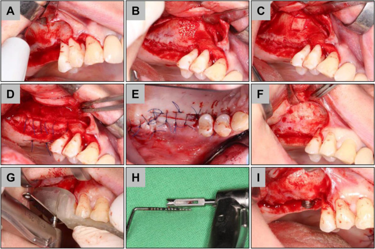
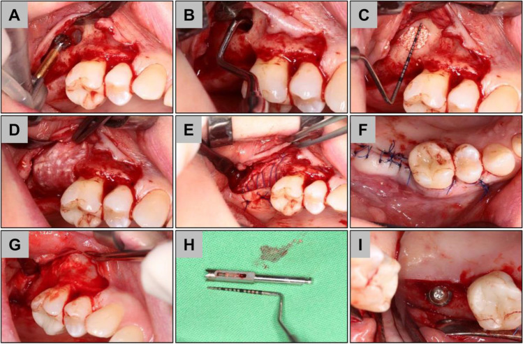
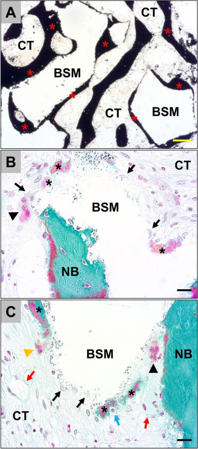
Forty patients were divided into two study groups. Both groups received a xenogeneic bone substitute material (BSM) during lateral sinus augmentation. In the bony wall group (BW), following piezosurgery, the retrieved bony wall was repositioned. In the collagen membrane group (CM), following rotary instrument preparation, collagen membrane coverage was applied. After 6 months, biopsies were taken to histologically analyze the percentage of BSM, connective tissue (CT), and newly formed bone (NFB) following both approaches.
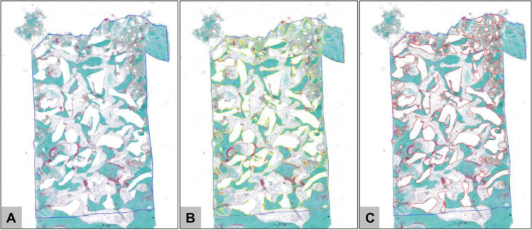
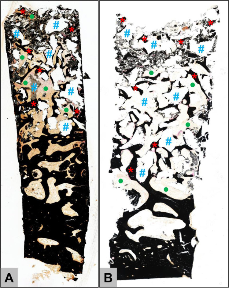
Forty implants were placed and 29 harvested biopsies could be evaluated. Duration of surgery, membrane perforations, and VAS were detected. Histomorphometrical analysis revealed comparable amounts of all analyzed parameters in both groups in descending order: CT (BW: 39.2 ± 9%, CM: 37,9 ± 8.5%) > BSM (BW: 32.9 ± 6.3%, CM: 31.8 ± 8.8%) > NB (BW: 27.8 ± 11.2%, CM: 30.3 ± 4.5%).
The results of the present study show that the closure of the access window by means of the retrieved bony wall or a native collagen membrane led to comparable bone augmentation results.
clinicaltrials.gov NCT04811768.
Lateral maxillary sinus augmentation with the application of a xenogeneic BSM in combination with a native collagen membrane for bony window coverage represents a reliable method for surgical reconstruction of the posterior maxilla. Piezosurgery with bony window repositioning delivers comparable outcomes without membrane coverage.
本随机对照临床试验旨在研究在上颌窦侧壁增量术中,重新定位的骨壁或应用胶原膜是否会导致更理想的新硬组织形成。
将 40 名患者分为两组。两组均在侧壁窦增大时使用异种骨替代物(BSM)。在骨壁组(BW)中,使用骨切开术后,重新定位取出的骨壁。在胶原膜组(CM)中,在旋转器械准备后,应用胶原膜覆盖。6 个月后,取活检进行组织学分析,以比较两种方法后 BSM、结缔组织(CT)和新骨形成(NFB)的百分比。
共植入 40 个种植体,取 29 个活检进行评估。检测手术时间、膜穿孔和 VAS。组织形态计量学分析显示,两组的所有分析参数均相似,按降序排列:CT(BW:39.2±9%,CM:37.9±8.5%)>BSM(BW:32.9±6.3%,CM:31.8±8.8%)>NB(BW:27.8±11.2%,CM:30.3±4.5%)。
本研究结果表明,通过使用取出的骨壁或天然胶原膜关闭进入窗口导致了相似的骨增量结果。
clinicaltrials.gov NCT04811768。
在上颌窦侧壁增量术中应用异种 BSM 联合天然胶原膜覆盖骨窗是一种可靠的方法,可用于后上颌骨的手术重建。使用骨切开术重新定位骨窗而不覆盖膜可获得类似的结果。