Ioachim Gabriela, Warren Howard J M, Powers Jocelyn M, Staud Roland, Pukall Caroline F, Stroman Patrick W
Center for Neuroscience Studies, Queen's University, Kingston, ON, Canada.
Department of Medicine, University of Florida, Seffner, FL, United States.
Front Neurol. 2022 May 6;13:862976. doi: 10.3389/fneur.2022.862976. eCollection 2022.
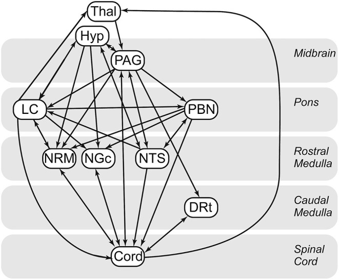
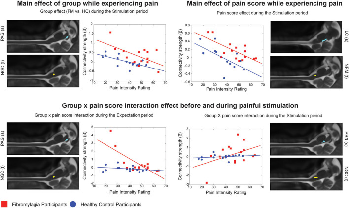
Chronic pain associated with fibromyalgia (FM) affects a large portion of the population but the underlying mechanisms leading to this altered pain are still poorly understood. Evidence suggests that FM involves altered neural processes in the central nervous system and neuroimaging methods such as functional magnetic resonance imaging (fMRI) are used to reveal these underlying alterations. While many fMRI studies of FM have been conducted in the brain, recent evidence shows that the changes in pain processing in FM may be linked to autonomic and homeostatic dysregulation, thus requiring further investigation in the brainstem and spinal cord. Functional magnetic resonance imaging data from 15 women with FM and 15 healthy controls were obtained in the cervical spinal cord and brainstem at 3 tesla using previously established methods. In order to investigate differences in pain processing in these groups, participants underwent trials in which they anticipated and received a predictable painful stimulus, randomly interleaved with trials with no stimulus. Differences in functional connectivity between the groups were investigated by means of structural equation modeling. The results demonstrate significant differences in brainstem/spinal cord network connectivity between the FM and control groups which also correlated with individual differences in pain responses. The regions involved in these differences in connectivity included the LC, hypothalamus, PAG, and PBN, which are known to be associated with autonomic homeostatic regulation, including fight or flight responses. This study extends our understanding of altered neural processes associated with FM and the important link between sensory and autonomic regulation systems in this disorder.
与纤维肌痛(FM)相关的慢性疼痛影响着很大一部分人群,但导致这种疼痛改变的潜在机制仍知之甚少。有证据表明,纤维肌痛涉及中枢神经系统神经过程的改变,功能磁共振成像(fMRI)等神经成像方法被用于揭示这些潜在的改变。虽然已经对纤维肌痛进行了许多大脑方面的fMRI研究,但最近的证据表明,纤维肌痛中疼痛处理的变化可能与自主神经和内稳态失调有关,因此需要在脑干和脊髓方面进行进一步研究。使用先前建立的方法,在3特斯拉的磁场下,获取了15名患有纤维肌痛的女性和15名健康对照者在颈脊髓和脑干的功能磁共振成像数据。为了研究这些组在疼痛处理方面的差异,参与者接受了试验,在试验中他们预期并接受了可预测的疼痛刺激,同时随机穿插无刺激的试验。通过结构方程模型研究了两组之间功能连接的差异。结果表明,纤维肌痛组和对照组在脑干/脊髓网络连接方面存在显著差异,这也与疼痛反应的个体差异相关。这些连接差异所涉及的区域包括蓝斑核(LC)、下丘脑、中脑导水管周围灰质(PAG)和臂旁核(PBN),这些区域已知与自主神经内稳态调节有关,包括战斗或逃跑反应。这项研究扩展了我们对与纤维肌痛相关的神经过程改变以及该疾病中感觉和自主调节系统之间重要联系的理解。