Wood Thomas R, Hildahl Kate, Helmbrecht Hawley, Corry Kylie A, Moralejo Daniel H, Kolnik Sarah E, Prater Katherine E, Juul Sandra E, Nance Elizabeth
Department of Pediatrics, Division of Neonatology University of Washington Seattle Washington USA.
Center on Human Development and Disability University of Washington Seattle Washington USA.
Bioeng Transl Med. 2021 Nov 23;7(2):e10265. doi: 10.1002/btm2.10265. eCollection 2022 May.
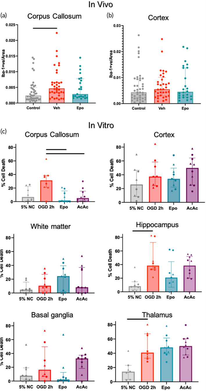
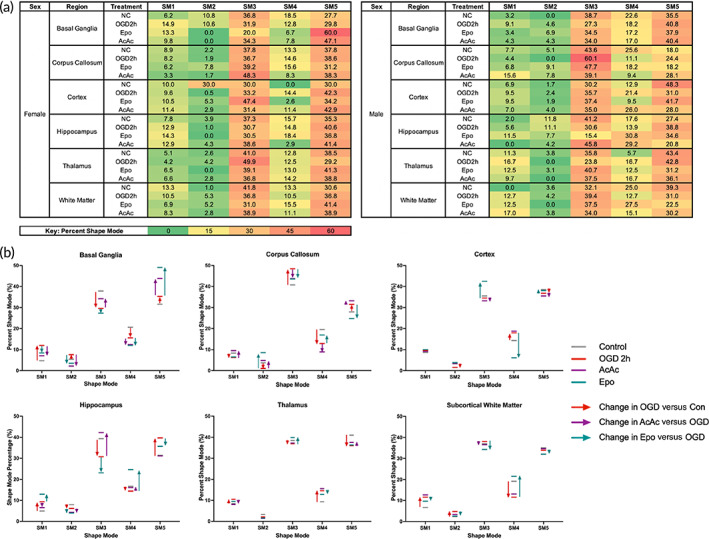
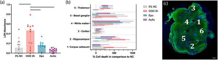
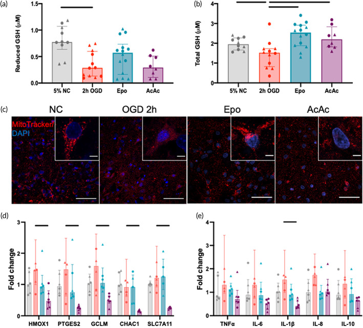
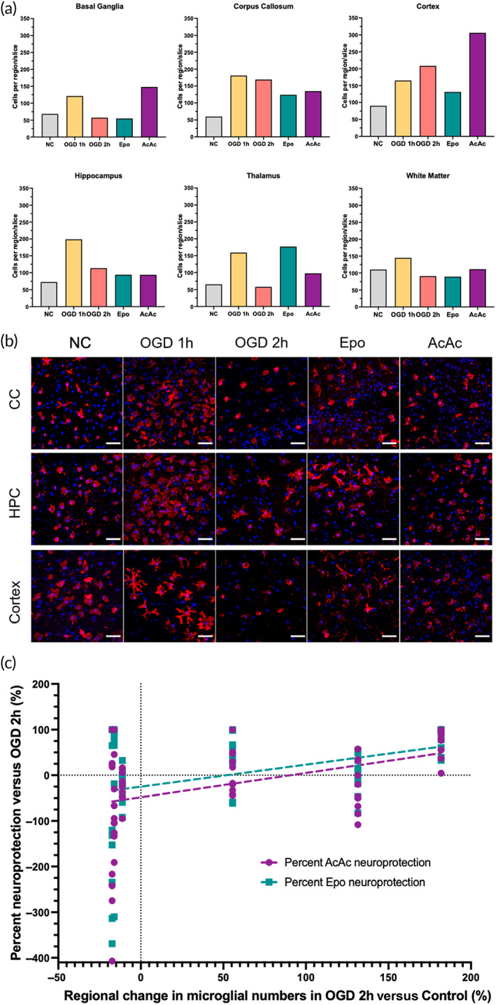
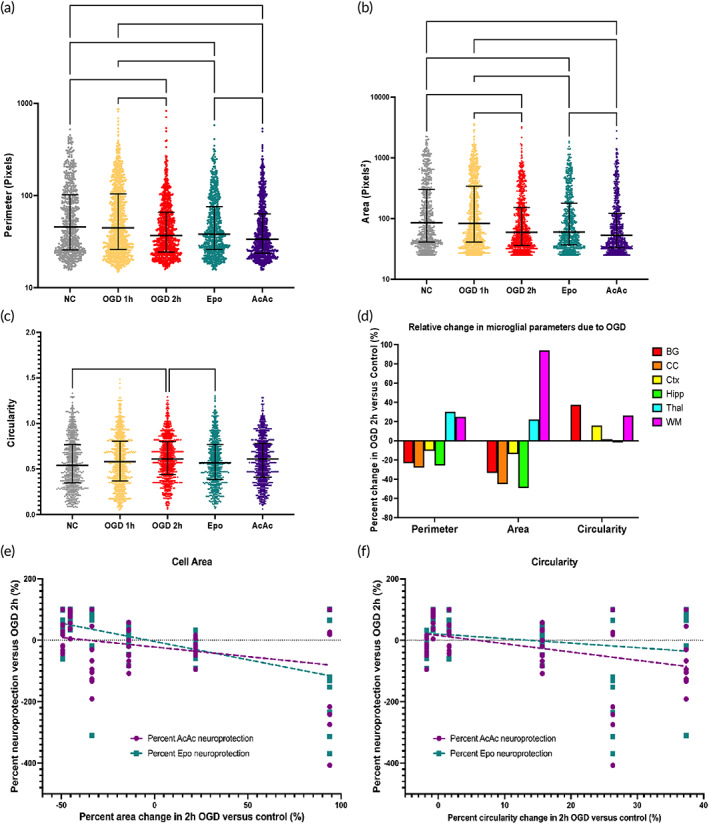
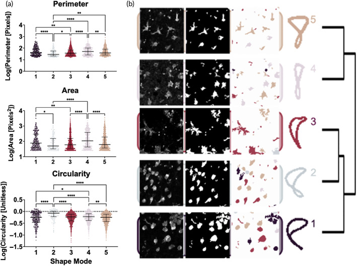
Organotypic brain slice models are an ideal technological platform to investigate therapeutic options for hypoxic-ischemic (HI) brain injury, a leading cause of morbidity and mortality in neonates. The brain exhibits regional differences in the response to HI injury in vivo. This can be modeled using organotypic brain slices, which maintain three-dimensional regional structures and reflect the regional differences in injury response. Here, we developed an organotypic whole hemisphere (OWH) slice culture model of HI injury using the gyrencephalic ferret brain at a developmental stage equivalent to a full-term human infant in order to better probe region-specific cellular responses to injury. Each slice encompassed the cortex, corpus callosum, subcortical white matter, hippocampus, basal ganglia, and thalamus. Regional responses to treatment with either erythropoietin (Epo) or the ketone body acetoacetate (AcAc) were highly heterogenous. While both treatments suppressed global injury responses and oxidative stress, significant neuroprotection was only seen in a subset of regions, with others displaying no response or potential exacerbation of injury. Similar regional heterogeneity was seen in the morphology and response of microglia to injury and treatment, which mirrored those seen after injury in vivo. Within each region, machine-learning-based classification of microglia morphological shifts in response to injury predicted the neuroprotective response to each therapy, with different morphologies associated with different treatment responses. This suggests that the ferret OWH slice culture model provides a platform for examining regional responses to injury in the gyrencephalic brain, as well as for screening combinations of therapeutics to provide global neuroprotection after injury.
器官型脑片模型是研究缺氧缺血性(HI)脑损伤治疗方案的理想技术平台,HI脑损伤是新生儿发病和死亡的主要原因。大脑在体内对HI损伤的反应存在区域差异。这可以通过器官型脑片来模拟,器官型脑片维持三维区域结构并反映损伤反应的区域差异。在此,我们使用与足月人类婴儿发育阶段相当的脑回状雪貂脑开发了一种HI损伤的器官型全脑(OWH)切片培养模型,以便更好地探究区域特异性细胞对损伤的反应。每片切片包含皮质、胼胝体、皮质下白质、海马体、基底神经节和丘脑。用促红细胞生成素(Epo)或酮体乙酰乙酸(AcAc)治疗后的区域反应高度异质性。虽然两种治疗都抑制了整体损伤反应和氧化应激,但仅在部分区域观察到显著的神经保护作用,其他区域则无反应或损伤可能加重。小胶质细胞对损伤和治疗的形态及反应也存在类似的区域异质性,这与体内损伤后观察到的情况相似。在每个区域内,基于机器学习对小胶质细胞对损伤反应的形态变化进行分类,可预测每种疗法的神经保护反应,不同的形态与不同的治疗反应相关。这表明雪貂OWH切片培养模型为研究脑回状脑对损伤的区域反应提供了一个平台,也为筛选治疗组合以在损伤后提供整体神经保护提供了平台。