McKenna Michael, Filteau Jeremy R, Butler Brendan, Sluis Kenneth, Chungyoun Michael, Schimek Nels, Nance Elizabeth
Department of Chemical Engineering, University of Washington, 105 Benson Hall, Box 351750, Seattle, WA, 98195-1750, USA.
Department of Chemistry, University of Washington, Seattle, WA, USA.
J Biol Eng. 2022 Jun 13;16(1):14. doi: 10.1186/s13036-022-00293-w.
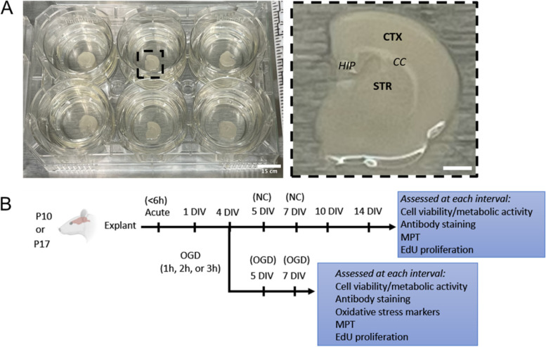
The brain extracellular environment is involved in many critical processes associated with neurodevelopment, neural function, and repair following injury. Organization of the extracellular matrix and properties of the extracellular space vary throughout development and across different brain regions, motivating the need for platforms that provide access to multiple brain regions at different stages of development. We demonstrate the utility of organotypic whole hemisphere brain slices as a platform to probe regional and developmental changes in the brain extracellular environment. We also leverage whole hemisphere brain slices to characterize the impact of cerebral ischemia on different regions of brain tissue.
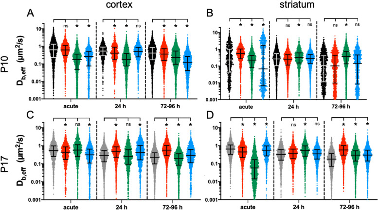
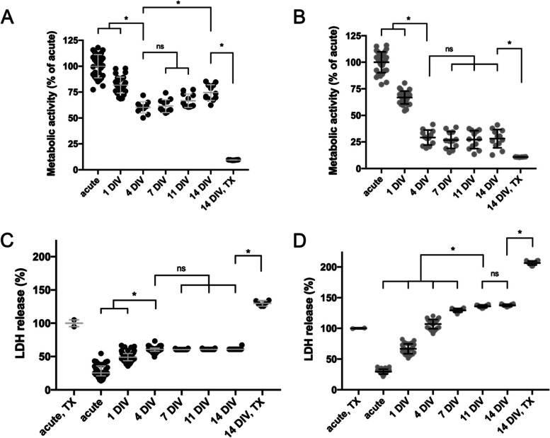
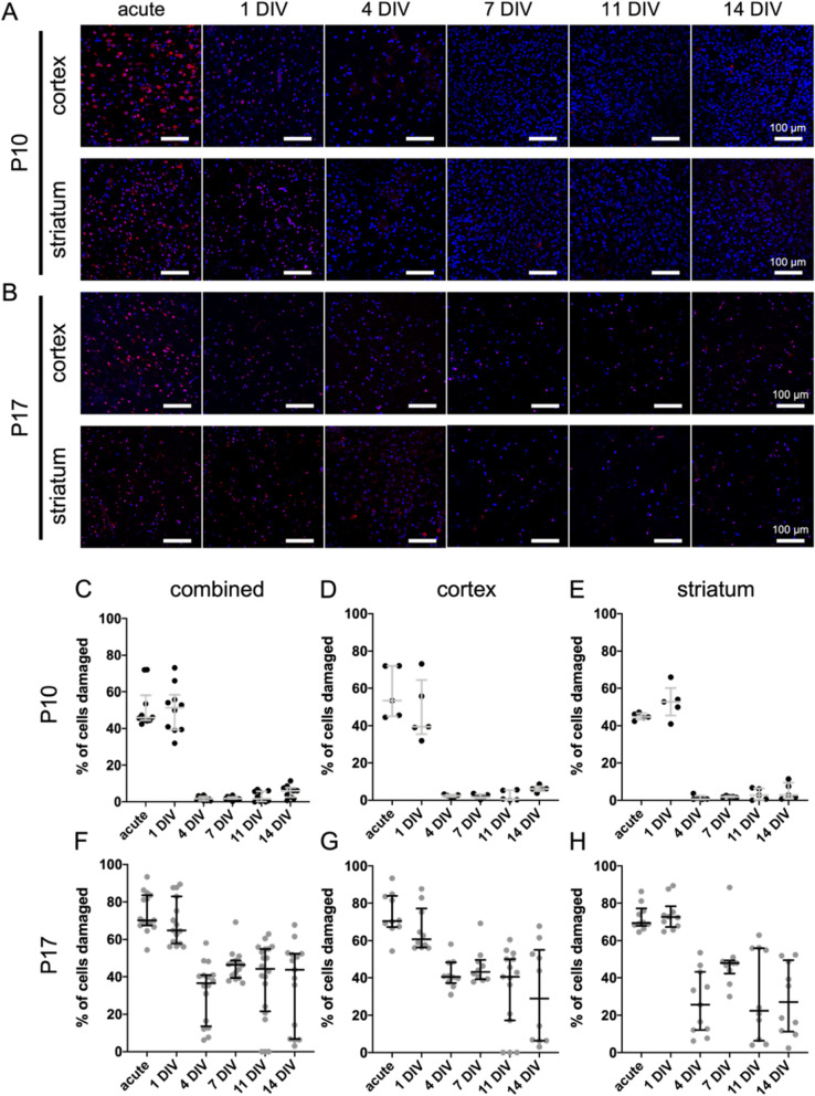
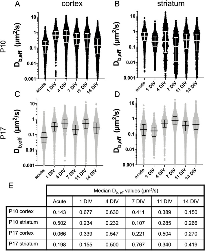
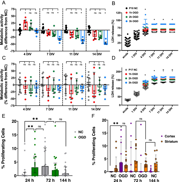
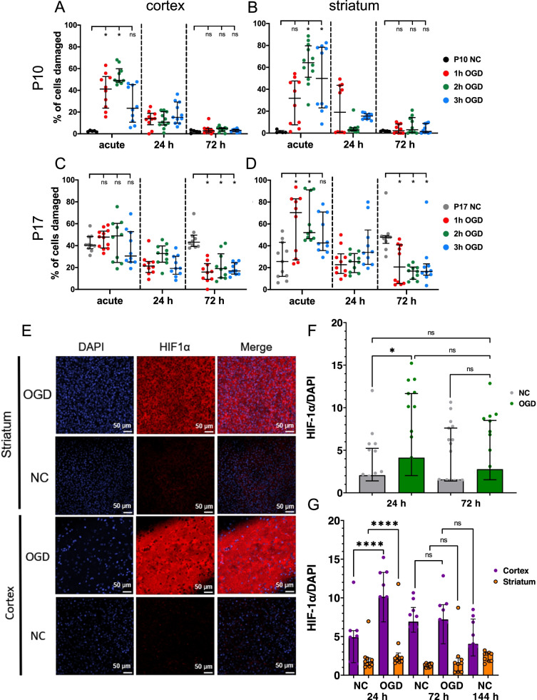
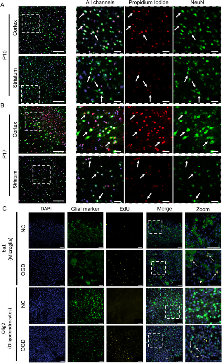
Whole hemisphere brain slices taken from postnatal (P) day 10 and P17 rats retained viable, metabolically active cells through 14 days in vitro (DIV). Oxygen-glucose-deprivation (OGD), used to model a cerebral ischemic event in vivo, resulted in reduced slice metabolic activity and elevated cell death, regardless of slice age. Slices from P10 and P17 brains showed an oligodendrocyte and microglia-driven proliferative response after OGD exposure, higher than the proliferative response seen in DIV-matched normal control slices. Multiple particle tracking in oxygen-glucose-deprived brain slices revealed that oxygen-glucose-deprivation impacts the extracellular environment of brain tissue differently depending on brain age and brain region. In most instances, the extracellular space was most difficult to navigate immediately following insult, then gradually provided less hindrance to extracellular nanoparticle diffusion as time progressed. However, changes in diffusion were not universal across all brain regions and ages.
We demonstrate whole hemisphere brain slices from P10 and P17 rats can be cultured up to two weeks in vitro. These brain slices provide a viable platform for studying both normal physiological processes and injury associated mechanisms with control over brain age and region. Ex vivo OGD impacted cortical and striatal brain tissue differently, aligning with preexisting data generated in in vivo models. These data motivate the need to account for both brain region and age when investigating mechanisms of injury and designing potential therapies for cerebral ischemia.
脑细胞外环境参与许多与神经发育、神经功能及损伤后修复相关的关键过程。细胞外基质的组织和细胞外空间的特性在整个发育过程以及不同脑区中各不相同,这促使人们需要能够在发育的不同阶段获取多个脑区的平台。我们展示了器官型全脑半球切片作为探究脑细胞外环境区域和发育变化的平台的实用性。我们还利用全脑半球切片来表征脑缺血对不同脑组织区域的影响。
取自出生后(P)第10天和第17天大鼠的全脑半球切片在体外培养14天(DIV)期间保留了存活的、具有代谢活性的细胞。用于模拟体内脑缺血事件的氧糖剥夺(OGD)导致切片代谢活性降低和细胞死亡增加,与切片年龄无关。来自P10和P17脑的切片在OGD暴露后显示出少突胶质细胞和小胶质细胞驱动的增殖反应,高于DIV匹配的正常对照切片中的增殖反应。对氧糖剥夺的脑切片进行多粒子追踪显示,氧糖剥夺对脑组织细胞外环境的影响因脑年龄和脑区而异。在大多数情况下,损伤后细胞外空间最初最难导航,然后随着时间的推移逐渐对细胞外纳米颗粒扩散的阻碍减少。然而,扩散变化在所有脑区和年龄中并非普遍存在。
我们证明取自P10和P17大鼠的全脑半球切片可在体外培养长达两周。这些脑切片为研究正常生理过程和损伤相关机制提供了一个可行的平台,可控制脑年龄和脑区。体外OGD对皮质和纹状体脑组织的影响不同,这与体内模型中已有的数据一致。这些数据表明,在研究损伤机制和设计脑缺血潜在治疗方法时,需要考虑脑区和年龄。