Department of Dermatology, STDs & Andrology, Misr University for Science & Technology (MUST), Giza, Egypt.
Department of Dermatology, STDs & Andrology, Faculty of Medicine, Fayoum University, Fayoum, Egypt.
Int Wound J. 2023 Jan;20(1):38-45. doi: 10.1111/iwj.13834. Epub 2022 May 23.
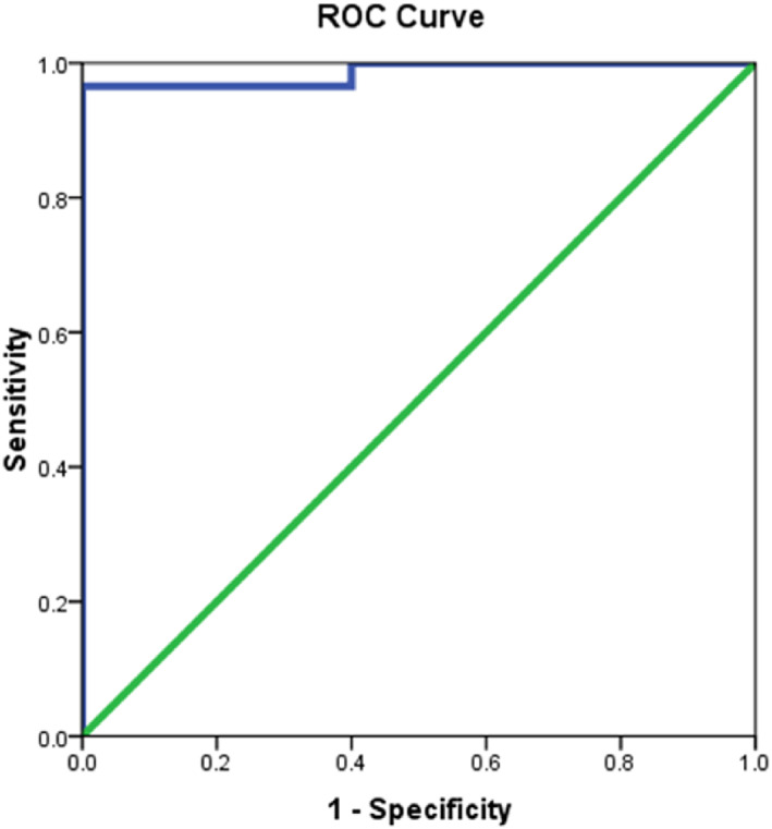
The pathophysiology of keloid formation is unknown, however, macrophages are thought to play a role in keloid formation. Understanding the mechanism(s) of keloid development might be crucial in developing a new treatment regimen for keloids. The aim of this study was to understand possible status of M1 and M2 type macrophages in the pathogenesis of keloid. Thirty cases of Keloid tissues were selected according to our inclusion and exclusion criteria, as well as 30 normal scars, were enrolled in our study as a control group. An excisional biopsy was harvested and ELISA was done on keloid tissue and normal scar samples, with CD68, the surface marker for M1 and CD163 representing M2. The results revealed the low expression of M1 (CD68) in keloid tissue meanwhile high levels of M1 were detected in normal scars. We also detected that higher tissue expression of M2 (CD163) was significantly associated with keloid cases when compared to low M2 expression in the control group. An important finding that was discovered during our study is that the M1 and M2 are significant predictors of keloid. Every increase of 1 ng/mL in M1 decreases the risk of keloid by 0.99 while every increase of one unit in M2 increases the risk of keloid by 2.01. This study concluded that the keloid formation could be a result of an abnormal response to tissue injury where there is an excessive entry of inflammatory cells into the wound, including macrophages and that the keloid incidence might be related to a decrease in M1 and an increase in M2.
瘢痕疙瘩形成的病理生理学尚不清楚,然而,巨噬细胞被认为在瘢痕疙瘩形成中起作用。了解瘢痕疙瘩发展的机制对于开发新的瘢痕疙瘩治疗方案可能至关重要。本研究旨在了解 M1 和 M2 型巨噬细胞在瘢痕疙瘩发病机制中的可能状态。根据纳入和排除标准,选择了 30 例瘢痕疙瘩组织作为研究对象,并纳入了 30 例正常瘢痕作为对照组。进行了切除活检,并对瘢痕疙瘩组织和正常瘢痕样本进行了 ELISA 检测,使用 CD68(M1 的表面标志物)和 CD163 代表 M2。结果显示,M1(CD68)在瘢痕疙瘩组织中的表达较低,而在正常瘢痕中则检测到高水平的 M1。我们还发现,与对照组中低水平的 M2 相比,M2(CD163)在组织中的高表达与瘢痕疙瘩病例显著相关。在我们的研究中发现的一个重要发现是,M1 和 M2 是瘢痕疙瘩的重要预测因子。M1 每增加 1ng/mL,瘢痕疙瘩的风险就会降低 0.99,而 M2 每增加一个单位,瘢痕疙瘩的风险就会增加 2.01。本研究得出结论,瘢痕疙瘩的形成可能是组织损伤的异常反应的结果,其中炎症细胞(包括巨噬细胞)过度进入伤口,而瘢痕疙瘩的发生率可能与 M1 的减少和 M2 的增加有关。