Ogawa Rei
Department of Plastic, Reconstructive and Aesthetic Surgery, Nippon Medical School Hospital, Tokyo 113-8603, Japan.
Int J Mol Sci. 2017 Mar 10;18(3):606. doi: 10.3390/ijms18030606.
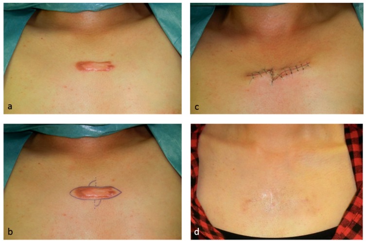
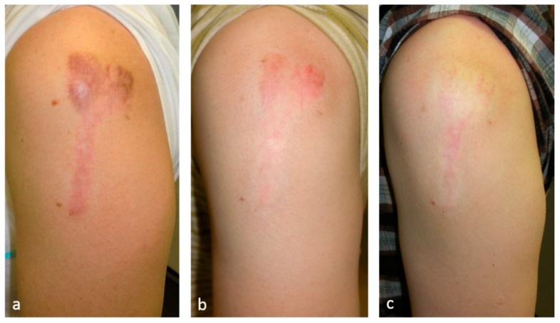
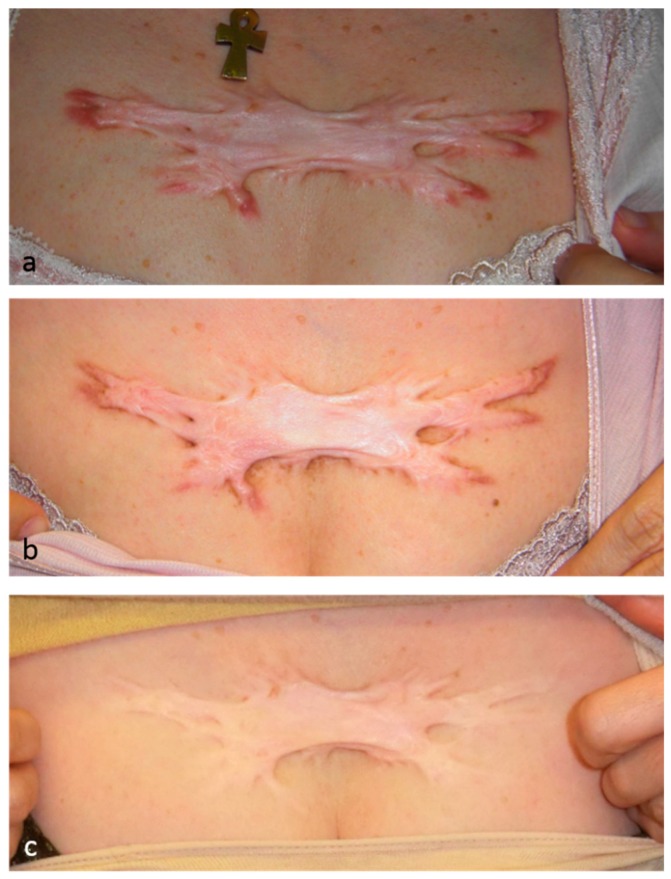
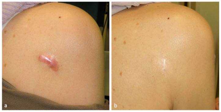
Keloids and hypertrophic scars are caused by cutaneous injury and irritation, including trauma, insect bite, burn, surgery, vaccination, skin piercing, acne, folliculitis, chicken pox, and herpes zoster infection. Notably, superficial injuries that do not reach the reticular dermis never cause keloidal and hypertrophic scarring. This suggests that these pathological scars are due to injury to this skin layer and the subsequent aberrant wound healing therein. The latter is characterized by continuous and histologically localized inflammation. As a result, the reticular layer of keloids and hypertrophic scars contains inflammatory cells, increased numbers of fibroblasts, newly formed blood vessels, and collagen deposits. Moreover, proinflammatory factors, such as interleukin (IL)-1α, IL-1β, IL-6, and tumor necrosis factor-α are upregulated in keloid tissues, which suggests that, in patients with keloids, proinflammatory genes in the skin are sensitive to trauma. This may promote chronic inflammation, which in turn may cause the invasive growth of keloids. In addition, the upregulation of proinflammatory factors in pathological scars suggests that, rather than being skin tumors, keloids and hypertrophic scars are inflammatory disorders of skin, specifically inflammatory disorders of the reticular dermis. Various external and internal post-wounding stimuli may promote reticular inflammation. The nature of these stimuli most likely shapes the characteristics, quantity, and course of keloids and hypertrophic scars. Specifically, it is likely that the intensity, frequency, and duration of these stimuli determine how quickly the scars appear, the direction and speed of growth, and the intensity of symptoms. These proinflammatory stimuli include a variety of local, systemic, and genetic factors. These observations together suggest that the clinical differences between keloids and hypertrophic scars merely reflect differences in the intensity, frequency, and duration of the inflammation of the reticular dermis. At present, physicians cannot (or at least find it very difficult to) control systemic and genetic risk factors of keloids and hypertrophic scars. However, they can use a number of treatment modalities that all, interestingly, act by reducing inflammation. They include corticosteroid injection/tape/ointment, radiotherapy, cryotherapy, compression therapy, stabilization therapy, 5-fluorouracil (5-FU) therapy, and surgical methods that reduce skin tension.
瘢痕疙瘩和增生性瘢痕是由皮肤损伤和刺激引起的,包括创伤、昆虫叮咬、烧伤、手术、接种疫苗、皮肤穿刺、痤疮、毛囊炎、水痘和带状疱疹感染。值得注意的是,未累及网状真皮层的浅表损伤不会导致瘢痕疙瘩和增生性瘢痕形成。这表明这些病理性瘢痕是由于该皮肤层的损伤以及随后在其中发生的异常伤口愈合所致。后者的特征是持续且在组织学上局限的炎症。因此,瘢痕疙瘩和增生性瘢痕的网状层含有炎症细胞、成纤维细胞数量增加、新形成的血管和胶原沉积。此外,促炎因子,如白细胞介素(IL)-1α、IL-1β、IL-6和肿瘤坏死因子-α在瘢痕疙瘩组织中上调,这表明在瘢痕疙瘩患者中,皮肤中的促炎基因对创伤敏感。这可能会促进慢性炎症,进而可能导致瘢痕疙瘩的侵袭性生长。此外,病理性瘢痕中促炎因子的上调表明,瘢痕疙瘩和增生性瘢痕不是皮肤肿瘤,而是皮肤的炎症性疾病,特别是网状真皮层的炎症性疾病。各种外部和内部的创伤后刺激可能会促进网状炎症。这些刺激的性质很可能决定了瘢痕疙瘩和增生性瘢痕的特征、数量和病程。具体而言,这些刺激的强度、频率和持续时间可能决定瘢痕出现的速度、生长方向和速度以及症状的严重程度。这些促炎刺激包括多种局部、全身和遗传因素。这些观察结果共同表明,瘢痕疙瘩和增生性瘢痕之间的临床差异仅仅反映了网状真皮层炎症的强度、频率和持续时间的差异。目前,医生无法(或至少发现非常困难)控制瘢痕疙瘩和增生性瘢痕的全身和遗传风险因素。然而,他们可以使用多种治疗方法,有趣的是,所有这些方法都通过减轻炎症起作用。它们包括皮质类固醇注射/贴剂/软膏、放射治疗、冷冻治疗、压迫治疗、稳定治疗、5-氟尿嘧啶(5-FU)治疗以及减轻皮肤张力的手术方法。