Institute of Medical Microbiology, Virology and Hygiene, University Medical Center Hamburg Eppendorf, Hamburg, Germany.
PLoS Pathog. 2022 May 23;18(5):e1010251. doi: 10.1371/journal.ppat.1010251. eCollection 2022 May.
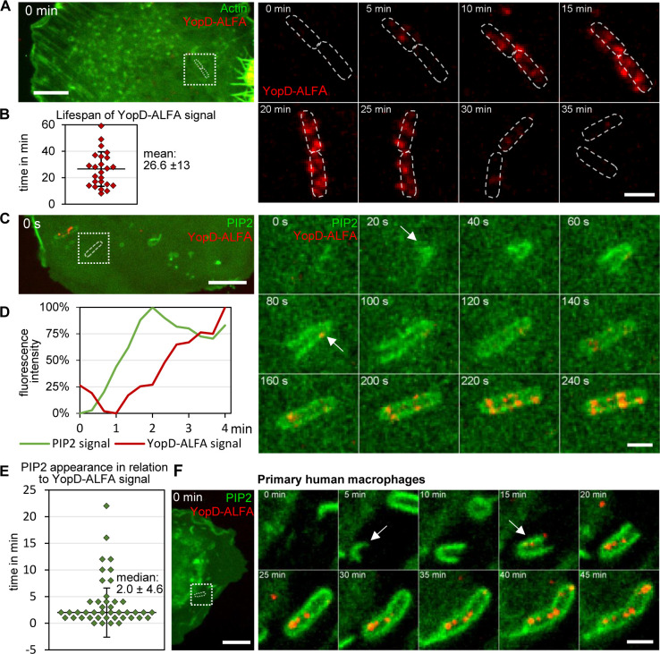
Yersinia enterocolitica employs a type three secretion system (T3SS) to translocate immunosuppressive effector proteins into host cells. To this end, the T3SS assembles a translocon/pore complex composed of the translocator proteins YopB and YopD in host cell membranes serving as an entry port for the effectors. The translocon is formed in a Yersinia-containing pre-phagosomal compartment that is connected to the extracellular space. As the phagosome matures, the translocon and the membrane damage it causes are recognized by the cell-autonomous immune system. We infected cells in the presence of fluorophore-labeled ALFA-tag-binding nanobodies with a Y. enterocolitica strain expressing YopD labeled with an ALFA-tag. Thereby we could record the integration of YopD into translocons and its intracellular fate in living host cells. YopD was integrated into translocons around 2 min after uptake of the bacteria into a phosphatidylinositol-4,5-bisphosphate enriched pre-phagosomal compartment and remained there for 27 min on average. Damaging of the phagosomal membrane as visualized with recruitment of GFP-tagged galectin-3 occurred in the mean around 14 min after translocon formation. Shortly after recruitment of galectin-3, guanylate-binding protein 1 (GBP-1) was recruited to phagosomes, which was accompanied by a decrease in the signal intensity of translocons, suggesting their degradation or disassembly. In sum, we were able for the first time to film the spatiotemporal dynamics of Yersinia T3SS translocon formation and degradation and its sensing by components of the cell-autonomous immune system.
耶尔森氏菌利用 III 型分泌系统(T3SS)将免疫抑制效应蛋白转运到宿主细胞中。为此,T3SS 组装了一个转位器/孔复合物,该复合物由宿主细胞膜中的转位蛋白 YopB 和 YopD 组成,作为效应蛋白的进入口。转位器是在含有耶尔森氏菌的前吞噬小体隔室中形成的,该隔室与细胞外空间相连。随着吞噬体的成熟,细胞自主免疫系统会识别转位器及其造成的膜损伤。我们在存在荧光标记的 ALFA 标签结合纳米抗体的情况下感染细胞,用表达带有 ALFA 标签的 YopD 的耶尔森氏菌菌株感染细胞。由此,我们可以记录 YopD 整合到转位器中的情况及其在活宿主细胞中的细胞内命运。在将细菌摄取到富含磷脂酰肌醇-4,5-二磷酸的前吞噬小体隔室中约 2 分钟后,YopD 被整合到转位器中,平均平均停留 27 分钟。在用 GFP 标记的半乳糖凝集素-3 招募来可视化吞噬体膜的损伤大约在转位器形成后 14 分钟左右发生。在半乳糖凝集素-3 招募后不久,鸟苷酸结合蛋白 1(GBP-1)被招募到吞噬体中,这伴随着转位器信号强度的降低,表明它们的降解或解体。总之,我们首次能够拍摄耶尔森氏菌 T3SS 转位器形成和降解的时空动态及其被细胞自主免疫系统成分的感应。