• 文献检索
  • 文档翻译
  • 深度研究
  • 学术资讯
  • Suppr Zotero 插件Zotero 插件
  • 邀请有礼
  • 套餐&价格
  • 历史记录
应用&插件
Suppr Zotero 插件Zotero 插件浏览器插件Mac 客户端Windows 客户端微信小程序
定价
高级版会员购买积分包购买API积分包
服务
文献检索文档翻译深度研究API 文档MCP 服务
关于我们
关于 Suppr公司介绍联系我们用户协议隐私条款
关注我们

Suppr 超能文献

核心技术专利:CN118964589B侵权必究
粤ICP备2023148730 号-1Suppr @ 2026

文献检索

告别复杂PubMed语法,用中文像聊天一样搜索,搜遍4000万医学文献。AI智能推荐,让科研检索更轻松。

立即免费搜索

文件翻译

保留排版,准确专业,支持PDF/Word/PPT等文件格式,支持 12+语言互译。

免费翻译文档

深度研究

AI帮你快速写综述,25分钟生成高质量综述,智能提取关键信息,辅助科研写作。

立即免费体验

严重小血管病患者的磁敏感加权成像上血管簇征的发生率及意义。

Prevalence and Significance of the Vessel-Cluster Sign on Susceptibility-Weighted Imaging in Patients With Severe Small Vessel Disease.

机构信息

From the Comprehensive Stroke Center (S.R.), Department of Neuroscience, Hospital Clinic, University of Barcelona; August Pi i Sunyer Biomedical Research Institute (IDIBAPS)(S.R.), Barcelona, Spain; Centre for Clinical Brain Sciences (E.C., M.S.S., M.T., F.C., G.B., D.J.G., F.D., I.H., J.M.W.), UK Dementia Research Institute, University of Edinburgh, United Kingdom; Institute for Stroke and Dementia Research (A.K., M. Dichgans), University Hospital, LMU Munich; Department of Radiology (M.I.),Ludwig-Maximilians-University Hospital Munich, Germany; Department of Neurology (D.K., J.S., R.v.O.), CARIM-School for Cardiovascular Diseases Maastricht University Medical Center+, Maastricht,; Department of Radiology & Nuclear Medicine (W.H.B.), School for Mental Health & Neuroscience and School for Cardiovascular Diseases, Maastricht University Medical Centre, Netherlands; Institute for Stroke and Dementia Research (ISD) (M. Duering), University Hospital, LMU Munich, Germany; Medical Image Analysis Center (MIAC AG) and Department of Biomedical Engineering (M. Duering), University of Basel, Switzerland; Munich Cluster for Systems Neurology (SyNergy) (M. Dichgans); and German Center for Neurodegenerative Diseases (DZNE) (M. Dichgans), Munich, Germany.

出版信息

Neurology. 2022 Aug 1;99(5):e440-e452. doi: 10.1212/WNL.0000000000200614.

DOI:10.1212/WNL.0000000000200614
PMID:35606147
原文链接:https://pmc.ncbi.nlm.nih.gov/articles/PMC9421604/
Abstract

BACKGROUND AND OBJECTIVES

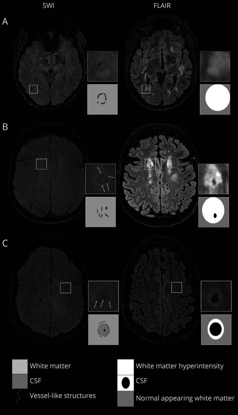
Magnetic resonance susceptibility-weighted imaging (SWI) can identify small brain blood vessels that contain deoxygenated blood due to its induced magnetic field disturbance. We observed focal clusters of possible dilated small vessels on SWI in white matter in severe small vessel disease (SVD). We assessed their prevalence, associations with SVD lesions, and vascular reactivity in patients with sporadic SVD and in patients with cerebral autosomal dominant arteriopathy with subcortical infarcts and leukoencephalopathy (CADASIL).

METHODS

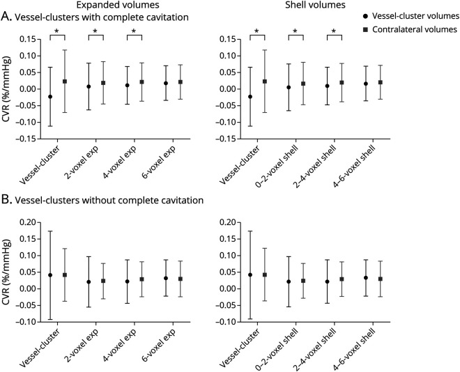
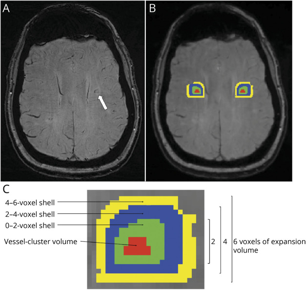
Secondary cross-sectional analysis of a prospective multicenter observational study of patients with either sporadic SVD or CADASIL (INVESTIGATE-SVD) studied with 3 Tesla MRI including blood-oxygen-level-dependent MRI cerebrovascular reactivity (CVR). Two independent raters evaluated SWI sequences to identify "vessel-clusters" in white matter as focal low-signal dots/lines with small vessel appearance (interrater agreement, kappa statistic = 0.66). We assessed per-patient and per-cluster associations with SVD lesion type and severity on structural MRI sequences. We also assessed CVR within and at 2-voxel concentric intervals around the vessel-clusters using contralateral volumes as a reference.

RESULTS

Among the 77 patients enrolled, 76 had usable SWI sequences, 45 with sporadic SVD (mean age 64 years [SD 11], 26 men [58%]) and 31 with CADASIL (53 years [11], 15 men [48%]). We identified 94 vessel-clusters in 36 of the 76 patients (15/45 sporadic SVD, 21/31 CADASIL). In covariate-adjusted analysis, patients with vessel-clusters had more lacunes (OR, 95% CI) (1.30, 1.05-1.62), higher white matter hyperintensity (WMH) volume (per-log10 increase, 1.92, 1.04-3.56), and lower CVR in normal appearing white matter (per %/mm Hg, 0.77, 0.60-0.99), compared with patients without vessel-clusters. Fifty-seven of the 94 vessel-clusters (61%) corresponded to noncavitated or partially cavitated WMH on fluid-attenuated inversion recovery, and 37 of 94 (39%) to complete cavities. CVR magnitude was lower than in the corresponding contralateral volumes (mean difference [SD], , ) within vessel-cluster volumes (-0.00046 [0.00088], -3.021, 0.005) and in the surrounding volume expansion shells up to 4 voxels (-0.00011 [0.00031], -2.140, 0.039; -0.00010 [0.00027], -2.295, 0.028) in vessel-clusters with complete cavities, but not in vessel-clusters without complete cavitation.

DISCUSSION

Vessel-clusters might correspond to maximally dilated vessels in white matter that are approaching complete tissue injury and cavitation. The pathophysiologic significance of this new feature warrants further longitudinal investigation.

摘要

背景与目的

磁共振磁敏感加权成像(SWI)可以识别因磁场干扰而含有脱氧血液的小脑血管。我们在严重小血管疾病(SVD)的脑白质中观察到 SWI 上可能存在的扩张小血管焦点簇。我们评估了它们的患病率,与 SVD 病变的相关性,以及散发性 SVD 患者和伴有皮质下梗死和白质脑病的常染色体显性脑动脉病(CADASIL)患者的血管反应性。

方法

对一项前瞻性多中心观察性研究的二级横断面分析,该研究纳入了 SVD 或 CADASIL 患者(INVESTIGATE-SVD),使用 3T MRI 进行研究,包括血氧水平依赖性 MRI 脑血管反应性(CVR)。两名独立的评分者评估 SWI 序列,以识别白质中的“血管簇”,即具有小血管外观的局灶性低信号点/线(组内一致性,kappa 统计量=0.66)。我们评估了每个患者和每个簇与结构 MRI 序列上 SVD 病变类型和严重程度的相关性。我们还使用对侧体积作为参考,在血管簇内和 2 个体素同心间隔内评估 CVR。

结果

在纳入的 77 名患者中,有 76 名患者的 SWI 序列可用,其中 45 名患有散发性 SVD(平均年龄 64 岁[标准差 11],26 名男性[58%]),31 名患有 CADASIL(53 岁[11],15 名男性[48%])。我们在 36 名患者中的 76 名患者中发现了 94 个血管簇(15/45 名散发性 SVD,21/31 名 CADASIL)。在调整协变量的分析中,有血管簇的患者腔隙数量更多(OR,95%CI)(1.30,1.05-1.62),脑白质高信号(WMH)体积更高(每对数增加 1.92,1.04-3.56),正常表现白质的 CVR 更低(每%/mmHg,0.77,0.60-0.99),与无血管簇的患者相比。94 个血管簇中的 57 个(61%)与液体衰减反转恢复的非空洞或部分空洞性 WMH 相对应,94 个中的 37 个(39%)与完全空洞相对应。与相应的对侧体积相比,血管簇内体积(平均差值[标准差],)内的 CVR 幅度较低(0.00046 [0.00088],-3.021,0.005),在 4 个体素的血管簇体积扩张壳内至 4 个体素(0.00011 [0.00031],-2.140,0.039;0.00010 [0.00027],-2.295,0.028)内,血管簇内存在完全空洞,但不存在不完全空洞。

讨论

血管簇可能对应于脑白质中最大扩张的血管,这些血管接近完全组织损伤和空洞化。这一新特征的病理生理意义值得进一步进行纵向研究。

https://cdn.ncbi.nlm.nih.gov/pmc/blobs/a1f5/9421604/481160b6fea5/WNL-2022-200666f5.jpg
https://cdn.ncbi.nlm.nih.gov/pmc/blobs/a1f5/9421604/504d44325e21/WNL-2022-200666f1.jpg
https://cdn.ncbi.nlm.nih.gov/pmc/blobs/a1f5/9421604/e5eb567bb6d5/WNL-2022-200666f2.jpg
https://cdn.ncbi.nlm.nih.gov/pmc/blobs/a1f5/9421604/98733feb450c/WNL-2022-200666f3.jpg
https://cdn.ncbi.nlm.nih.gov/pmc/blobs/a1f5/9421604/a02308c7d12f/WNL-2022-200666f4.jpg
https://cdn.ncbi.nlm.nih.gov/pmc/blobs/a1f5/9421604/481160b6fea5/WNL-2022-200666f5.jpg

相似文献

1
Prevalence and Significance of the Vessel-Cluster Sign on Susceptibility-Weighted Imaging in Patients With Severe Small Vessel Disease.严重小血管病患者的磁敏感加权成像上血管簇征的发生率及意义。
Neurology. 2022 Aug 1;99(5):e440-e452. doi: 10.1212/WNL.0000000000200614.
2
MarkVCID cerebral small vessel consortium: II. Neuroimaging protocols.马克 VCID 脑小血管联盟:二、神经影像学协议。
Alzheimers Dement. 2021 Apr;17(4):716-725. doi: 10.1002/alz.12216. Epub 2021 Jan 21.
3
Heterogeneity and Penumbra of White Matter Hyperintensities in Small Vessel Diseases Determined by Quantitative MRI.定量MRI确定的小血管疾病中白质高信号的异质性和半暗带
Stroke. 2025 Jan;56(1):128-137. doi: 10.1161/STROKEAHA.124.047910. Epub 2024 Dec 9.
4
Antithrombotic therapy to prevent cognitive decline in people with small vessel disease on neuroimaging but without dementia.抗血栓治疗预防神经影像学检查发现的小血管疾病但无痴呆的患者认知能力下降。
Cochrane Database Syst Rev. 2022 Jul 14;7(7):CD012269. doi: 10.1002/14651858.CD012269.pub2.
5
Cerebrovascular Function in Sporadic and Genetic Cerebral Small Vessel Disease.散发性和遗传性脑小血管病中的脑血管功能
Ann Neurol. 2025 Mar;97(3):483-498. doi: 10.1002/ana.27136. Epub 2024 Nov 18.
6
Arterial, Venous, and Cerebrospinal Fluid Flow and Pulsatility in Stroke-Related Cerebral Small Vessel Disease: A Longitudinal Analysis.与中风相关的脑小血管病中的动脉、静脉及脑脊液流动与搏动性:一项纵向分析
Stroke. 2025 Sep;56(9):2450-2463. doi: 10.1161/STROKEAHA.124.049103. Epub 2025 Jun 18.
7
Incidence of Diffusion-Weighted Imaging Lesions in Patients With Intracerebral Hemorrhage in the Acute and Subacute Time Periods.急性和亚急性期脑出血患者扩散加权成像病变的发生率
Neurology. 2025 Jul 22;105(2):e213837. doi: 10.1212/WNL.0000000000213837. Epub 2025 Jun 19.
8
Cerebral small vessel disease and renal function: systematic review and meta-analysis.脑小血管病与肾功能:系统评价与荟萃分析
Cerebrovasc Dis. 2015;39(1):39-52. doi: 10.1159/000369777. Epub 2014 Dec 24.
9
Enlarged perivascular spaces are associated with white matter injury, cognition and inflammation in cerebral autosomal dominant arteriopathy with subcortical infarcts and leukoencephalopathy.在伴有皮质下梗死和白质脑病的脑常染色体显性动脉病中,血管周围间隙扩大与白质损伤、认知及炎症相关。
Brain Commun. 2024 Mar 8;6(2):fcae071. doi: 10.1093/braincomms/fcae071. eCollection 2024.
10
Arterial Spin-Labeling MRI Identifies Abnormal Perfusion Metric at the Gray Matter/CSF Interface in Cerebral Small Vessel Disease.动脉自旋标记磁共振成像可识别脑小血管病中灰质/脑脊液界面的异常灌注指标。
AJNR Am J Neuroradiol. 2025 Jul 1;46(7):1345-1352. doi: 10.3174/ajnr.A8682.

引用本文的文献

1
Small vessel dysfunction at 7T MRI locally predicts white matter damage progression in CADASIL.7T磁共振成像显示的小血管功能障碍可局部预测伴有皮质下梗死和白质脑病的常染色体显性遗传性脑动脉病(CADASIL)中的白质损伤进展。
J Cereb Blood Flow Metab. 2025 Aug 20:271678X251369257. doi: 10.1177/0271678X251369257.
2
Retinal vascular reactivity is associated with white matter hyperintensities and dysfunctional cerebrovascular reactivity in cerebral small vessel disease.视网膜血管反应性与脑小血管病中的白质高信号及脑血管反应性异常有关。
J Cereb Blood Flow Metab. 2025 Aug 6:271678X251366079. doi: 10.1177/0271678X251366079.
3
"Chocolate Chip Sign" on Susceptibility-Weighted Imaging: A Novel Neuroimaging Biomarker for HTRA1-Related Cerebral Small Vessel Disease.

本文引用的文献

1
Imaging neurovascular, endothelial and structural integrity in preparation to treat small vessel diseases. The INVESTIGATE-SVDs study protocol. Part of the SVDs@Target project.为治疗小血管疾病而对神经血管、内皮和结构完整性进行成像。INVESTIGATE-SVDs研究方案。SVDs@Target项目的一部分。
Cereb Circ Cogn Behav. 2021 Jun 26;2:100020. doi: 10.1016/j.cccb.2021.100020. eCollection 2021.
2
Sex Differences in Cerebral Small Vessel Disease: A Systematic Review and Meta-Analysis.脑小血管病中的性别差异:一项系统评价与荟萃分析
Front Neurol. 2021 Oct 28;12:756887. doi: 10.3389/fneur.2021.756887. eCollection 2021.
3
磁敏感加权成像上的“巧克力片征”:一种用于与HTRA1相关的脑小血管病的新型神经影像学生物标志物。
Neurol Genet. 2025 Feb 26;11(2):e200237. doi: 10.1212/NXG.0000000000200237. eCollection 2025 Apr.
4
Cerebrovascular Function in Sporadic and Genetic Cerebral Small Vessel Disease.散发性和遗传性脑小血管病中的脑血管功能
Ann Neurol. 2025 Mar;97(3):483-498. doi: 10.1002/ana.27136. Epub 2024 Nov 18.
5
Prevalence and Clinical Implications of Hemosiderin Deposits in Recent Small Subcortical Infarcts.近期小皮质下梗死中铁红素沉积的患病率及其临床意义。
Neurology. 2024 Nov 26;103(10):e209973. doi: 10.1212/WNL.0000000000209973. Epub 2024 Oct 24.
6
Incident Infarcts in Patients With Stroke and Cerebral Small Vessel Disease: Frequency and Relation to Clinical Outcomes.脑小血管病患者脑卒中后梗死灶的发生频率及其与临床结局的关系。
Neurology. 2024 Sep 10;103(5):e209750. doi: 10.1212/WNL.0000000000209750. Epub 2024 Aug 19.
7
National and international collaborations to advance research into vascular contributions to cognitive decline.国内和国际合作推动对血管因素导致认知能力下降的研究。
Cereb Circ Cogn Behav. 2023 Dec 14;6:100195. doi: 10.1016/j.cccb.2023.100195. eCollection 2024.
8
Prevalence and Predictors of Hemorrhagic Foci on Long-term Follow-up MRI of Recent Single Subcortical Infarcts.近期单发性皮质下梗死长期随访MRI上出血灶的患病率及预测因素
Transl Stroke Res. 2025 Apr;16(2):410-420. doi: 10.1007/s12975-023-01224-7. Epub 2023 Dec 14.
9
Emerging frontiers of artificial intelligence and machine learning in ischemic stroke: a comprehensive investigation of state-of-the-art methodologies, clinical applications, and unraveling challenges.人工智能和机器学习在缺血性中风领域的新兴前沿:对前沿方法、临床应用及未解挑战的全面调查
EPMA J. 2023 Nov 2;14(4):645-661. doi: 10.1007/s13167-023-00343-3. eCollection 2023 Dec.
10
Deep learning based on susceptibility-weighted MR sequence for detecting cerebral microbleeds and classifying cerebral small vessel disease.基于磁敏感加权 MR 序列的深度学习用于检测脑微出血和分类脑小血管病。
Biomed Eng Online. 2023 Oct 17;22(1):99. doi: 10.1186/s12938-023-01164-1.
Cerebrovascular Reactivity Measurement Using Magnetic Resonance Imaging: A Systematic Review.
使用磁共振成像测量脑血管反应性:一项系统综述。
Front Physiol. 2021 Feb 25;12:643468. doi: 10.3389/fphys.2021.643468. eCollection 2021.
4
Susceptibility Vessel Sign in Deep Perforating Arteries in Patients with Recent Small Subcortical Infarcts.近期小皮质下梗死患者深穿支动脉易损斑块征。
J Stroke Cerebrovasc Dis. 2021 Jan;30(1):105415. doi: 10.1016/j.jstrokecerebrovasdis.2020.105415. Epub 2020 Nov 1.
5
Brain deep medullary veins on 3-T MRI in a population-based cohort.基于人群的队列研究中 3-T MRI 脑深部髓静脉成像。
J Cereb Blood Flow Metab. 2021 Mar;41(3):561-568. doi: 10.1177/0271678X20918467. Epub 2020 Apr 20.
6
Relationship Between Venules and Perivascular Spaces in Sporadic Small Vessel Diseases.散发性小血管病中小静脉与血管周围腔隙的关系。
Stroke. 2020 May;51(5):1503-1506. doi: 10.1161/STROKEAHA.120.029163. Epub 2020 Apr 8.
7
Decreased visible deep medullary veins is a novel imaging marker for cerebral small vessel disease.脑小血管病的新型影像学标志物:可见深部脑髓静脉减少。
Neurol Sci. 2020 Jun;41(6):1497-1506. doi: 10.1007/s10072-019-04203-9. Epub 2020 Jan 18.
8
Increased Diameters of the Internal Cerebral Veins and the Basal Veins of Rosenthal Are Associated with White Matter Hyperintensity Volume.Rosenthal 内静脉和基底静脉直径增宽与脑白质高信号体积相关。
AJNR Am J Neuroradiol. 2019 Oct;40(10):1712-1718. doi: 10.3174/ajnr.A6213. Epub 2019 Sep 12.
9
Small vessel disease: mechanisms and clinical implications.小血管疾病:机制与临床意义。
Lancet Neurol. 2019 Jul;18(7):684-696. doi: 10.1016/S1474-4422(19)30079-1. Epub 2019 May 13.
10
Elevated brain oxygen extraction fraction measured by MRI susceptibility relates to perfusion status in acute ischemic stroke.MRI 磁化率测量的脑氧摄取分数升高与急性缺血性卒中的灌注状态有关。
J Cereb Blood Flow Metab. 2020 Mar;40(3):539-551. doi: 10.1177/0271678X19827944. Epub 2019 Feb 7.